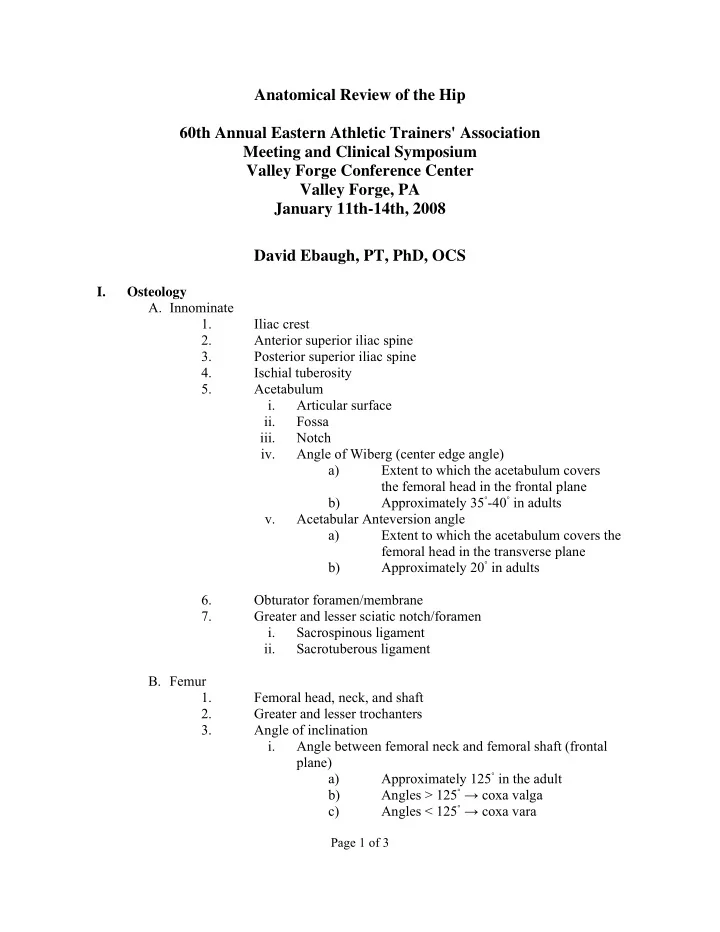

Anatomical Review of the Hip 60th Annual Eastern Athletic Trainers' Association Meeting and Clinical Symposium Valley Forge Conference Center Valley Forge, PA January 11th-14th, 2008 David Ebaugh, PT, PhD, OCS I. Osteology A. Innominate 1. Iliac crest 2. Anterior superior iliac spine 3. Posterior superior iliac spine 4. Ischial tuberosity 5. Acetabulum i. Articular surface ii. Fossa iii. Notch iv. Angle of Wiberg (center edge angle) a) Extent to which the acetabulum covers the femoral head in the frontal plane Approximately 35 ◦ -40 ◦ in adults b) v. Acetabular Anteversion angle a) Extent to which the acetabulum covers the femoral head in the transverse plane Approximately 20 ◦ in adults b) 6. Obturator foramen/membrane 7. Greater and lesser sciatic notch/foramen i. Sacrospinous ligament ii. Sacrotuberous ligament B. Femur 1. Femoral head, neck, and shaft 2. Greater and lesser trochanters 3. Angle of inclination i. Angle between femoral neck and femoral shaft (frontal plane) Approximately 125 ◦ in the adult a) Angles > 125 ◦ → coxa valga b) Angles < 125 ◦ → coxa vara c) Page 1 of 3
4. Torsion angle i. Angle between the shaft and neck of the femur (transverse plane) Approximately 10 ◦ -15 ◦ in the adult (normal a) anteversion) Angles > 15 ◦ → excessive anteversion b) Angles < 15 ◦ → retroversion c) II. Joint Capsule and Ligaments A. Fibrous portion of capsule; attachment sites B. Synovial portion of capsule; attachment sites 1. Femoral neck fractures and avascular necrosis C. Reinforcing ligaments 1. Iliofemoral (Y ligament of Bigelow) 2. Ischiofemoral 3. Pubofemoral III. Articular Surfaces and Acetabular labrum A. Acetabulum 1. Cartilage thickest superior and lateral 2. Ligament to head of femur (ligamentum teres) B. Femoral head 1. Cartilage thickest on medial central region C. Acetabular labrum 1. Fibrocartilagenous ring 2. Transverse acetabular ligament IV. Muscles A. Anterior 1. Iliopsoas 2. Sartorius 3. Rectus femoris 4. Tensor fascia lata B. Posterior 1. Gluteus maximus 2. Piriformis 3. Superior and inferior gemellus 4. Obturator internus 5. Quadratus femoris 6. Semimembranosus 7. Semitendinosus Page 2 of 3
8. Biceps femoris C. Lateral 1. Gluteus medius 2. Gluteus minimus 3. Tensor fascia lata and Iliotibial Band D. Medial 1. Pectineus 2. Adductor longus, brevis, and magnus 3. Gracilis 4. Obturator externus V. Neurovascular Structures A. Femoral nerve, artery, and vein 1. Deep femoral artery i. Medial and lateral femoral circumflex arteries ii. Perforating branches B. Lateral femoral cutaneous nerve C. Obturator nerve D. Superior and inferior gluteal nerve, artery, and vein E. Sciatic nerve F. Posterior femoral cutaneous nerve VI. Bursae A. Iliopsoas (liopectineal) 1. Between iliopsoas muscle and anterior joint capsule B. Trochanteric 1. Between gluteal muscles and greater trochanter Page 3 of 3
Recommend
More recommend