A Low Cost Approach to Specimen Level Imaging of Natural History - - PDF document
A Low Cost Approach to Specimen Level Imaging of Natural History - - PDF document
Downloaded from OSF: https://www.doi.org/10.31219/osf.io/dvmsh A Low Cost Approach to Specimen Level Imaging of Natural History Microscope Slides using a DSLR System E. Louise Allan 1 *, Benjamin W. Price 1 , Olha Shchedrina 1 , Steen Dupont 1 ,
Downloaded from OSF: https://www.doi.org/10.31219/osf.io/dvmsh A Low Cost Approach to Specimen Level Imaging of Natural History Microscope Slides using a DSLR System E. Louise Allan 1 *, Benjamin W. Price 1 , Olha Shchedrina 1 , Steen Dupont 1 , Laurence Livermore 1 , and Vince S. Smith 1 1 Natural History Museum, Cromwell Road, SW7 5BD, London *Corresponding author: louise.allan@nhm.ac.uk Abstract For specimen level imaging of microscope slides automated digital microscopy systems are widely used, however, these systems are not always suitable for non-standard slides such as those found in natural history collections. For these types of slides imaging will require the use of non-automated alternatives. This paper presents a low cost option for imaging non-standard slides, such as damaged or irregular slides, by using a DSLR camera with a specialised macro lens (5:1) mounted to a StackShot Macro Rail and a flashbox, known as the DSLR-StackShot system. There was no noticeable difference in the image resolution between the DSLR- StackShot system and 5x magnification using a high end automated slide scanner, such as the Axio Scan.Z1. The DSLR-StackShot system, while on partially automated, enables the required flexibility and manual control required when imaging slides of varying sizes, thicknesses, and preservation types, as well as slides that are damaged or in poor condition. Keywords Microscope slides, natural history collections, specimen level imaging, DSLR Background Digitisation of natural history specimens enables access to natural history collections for new audiences and improves research opportunities (Drew et al. 2017). In 2014 the Natural History Museum, London (NHM) embarked on an ambitious Digital Collections Programme (DCP) to digitise its collections, estimated to comprise 80 million specimens. One of the aims of the DCP is to develop digitisation workflows for each collection type. For slide mounted specimens two digitisation approaches can be utilised, one approach focuses on capturing an overview image of the slide and its associated labels (Heerlien et al. 2015; Allan et al. in review; Summerfield et al. in prep), while the other focuses on specialised imaging of the slide mounted material (Rojo et al. 2006; Musson et al. 2015; Summerfield et al. in prep). 1
Downloaded from OSF: https://www.doi.org/10.31219/osf.io/dvmsh Overview imaging of microscope slides can be carried out using non-complex systems and can be used for mass digitisation of collections to extract specimen data (Heerlien et al. 2015; Allan et al. in review). Specimen level imaging on the other hand requires digital microscopy systems (Rojo et al. 2006). Automated slide digitisation systems, designed for higher resolution imaging of standardised material, have existed for over a decade but have been confined to medical slides with no other large-scale digitisation projects of natural history slides known to us (Rojo et al. 2006, Weinstein et al. 2009, Dietrich et al. 2012). While there have been several pilot projects that have used specially modified histology slide scanners adapted for natural history specimens, they cannot accommodate damaged slides, or slides with non-standard thickness or length (Musson et al. 2015; Summerfield et al. in prep) - issues that can be frequent in natural history collections (Figure 1). Figure 1. Examples of natural history microscope slides that are damaged or non-standard in size and mountant thickness. We estimate that the NHM holds 2.4 million microscope slides in its collection. These slides are distributed across diverse curatorial groups (e.g. botany, entomology, mineralogy, palaeontology and zoology), with each group having its own distinct slide preparation technique and standards. In 2015 the Museum’s DCP conducted a pilot project to digitise a variety of slide mounted material using a specially modified histology slide scanner, ZEISS Axio Scan.Z1 (Summerfield et al in prep). The Axio Scan is a highly automated system that can image up to a 100 slides per run (Figure 2); however, it is designed to image standardised slides and thus adaptations were required to enable digitisation of natural history slides. Some slides, however, could not be imaged using the Axio Scan i.e. damaged slides or those with thick mountants, and had to be imaged using a non-automated system, such as the Axio Zoom.V16 (Summerfield et al. in prep). 2
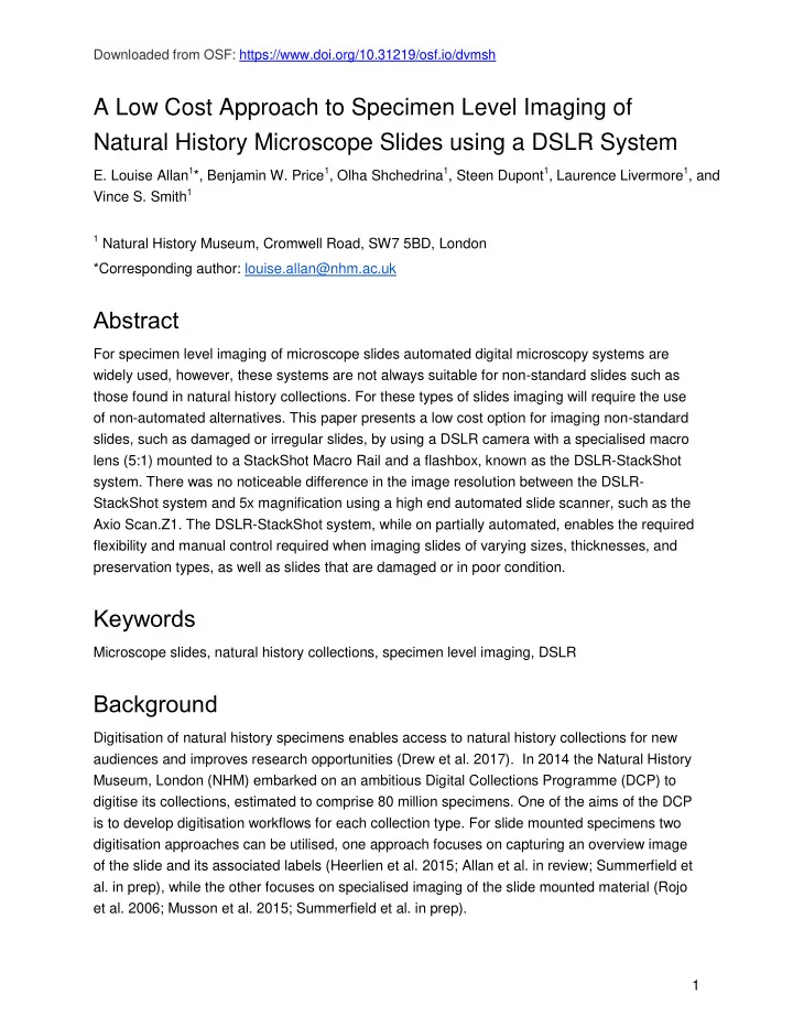
Downloaded from OSF: https://www.doi.org/10.31219/osf.io/dvmsh Figure 2. Axio Scan.Z1 is an automated slide scanner that uses holders to move the slides into position for imaging and can image up to 100 slides per run. In 2017 the DCP digitised the microscope slide collection of Phthiraptera (Allan et al. in review), of which one of the aims was to specimen level image a representative of each species, focusing on type material where present. As expected a proportion of slides were unsuitable for Axio Scan and were imaged using a Canon EOS 5DS R with a MP-E 65mm lens, StackShot Macro Rail system (Cognisys Inc.), and a custom flashbox (Figure 3). This customised setup enabled us to adapt our existing whole slide imaging setup, described in Allan et al. (in review), thus creating a low cost alternative for specimen level imaging of microscope slides. Figure 3. DSLR-StackShot system uses a DSLR camera vertically mounted to a StackShot Macro Rail with a custom flashbox and microscope stage. 3
Downloaded from OSF: https://www.doi.org/10.31219/osf.io/dvmsh DSLR - StackShot System Hardware We used a DSLR camera (Canon EOS 5DS R) with a specialist macro lens (5:1; MP-E 65mm), which is capable of obtaining a comparable image resolution to the 5x magnification obtained using the Axio Scan (Figure 4). When combined with a StackShot Macro Rail, the system is capable of taking extended depth of field images. The slide is illuminated from below using a flash that is housed in a custom built box that is fitted with a microscope stage to enable fine scale movement of the slide (Figure 3). (a) (b) Figure 4 . Image resolution comparison between (a) the Axio Scan (5x magnification) and (b) the DSLR- StackShot system. Specimen: human head louse. Left column: overview of the specimen; right column: full size resolution. 4
Downloaded from OSF: https://www.doi.org/10.31219/osf.io/dvmsh Software Images were captured using Helicon Remote v.3.8.4 W (https://www.heliconsoft.com/heliconsoft-products/helicon-remote/), while the integrated StackShot macro rail system was used to set the focal range and interval between images, thus enabling automated stack image capture. The camera mode was set to manual with the aperture set between f 1/5.6; ISO 100 and shutter speed 1/200 sec. The light source consisted of a flash using a power setting between 1/16 and 1/32. Each set of images was saved to a new folder, which was renamed with the specimen’s unique identi fier (UID) i.e. barcode number. At the end of the day the images were stacked as a batch using Helicon Focus v.6.7.1 (https://www.heliconsoft.com/heliconsoft-products/helicon-focus/). The stacked images were then renamed in bulk with their corresponding folder name (UID) using Bulk Rename Utility v.3.0.0.1 (https://www.bulkrenameutility.co.uk/Download.php), and the scale bar was stamped using an ImageJ v.1.52h macro within Fiji (https://imagej.net/Fiji/Downloads). Unlike the Axio Scan the addition of scale bars for the DSLR-StackShot system is a manual step and requires new scale bars to be calculated if the magnification changes. To minimise this manual step, images were captured at the same magnification i.e. at the full lens magnification, thus facilitating batch processing using an ImageJ macro. If the specimen was larger than the field of view then a lower magnification, and new scale bar, was required. When possible these specimens were imaged in batches. Adaptations are currently being investigated for more automated ways to identify the lens magnification and associated scale. Specimen Level Imaging of Natural History Slides During the two DCP slide digitisation projects a subset of slides were selected for specimen level imaging using the Axio Scan. As the Axio Scan is optimised for imaging uniform slides that are adequately stained, adaptations were required in order to image our natural history slides. Some slides, however, were unsuitable for imaging using this automated system, despite the adaptations, and were imaged using the DSLR_StackShot system. Below we list a number of issues common to natural history slides that hindered the use of the automated scanner. 1. Slide suitability Variation in size and thickness of slides is common among natural history slides. In order to image larger slides using Axio Scan, ZEISS created a variety of holders that could accommodate different sized slides; however, some slides were too irregular or delicate to be placed in these holders. With the DSLR-StackShot system, as the slide can be place ‘freely’ on the flashbox within the field of view it can accommodate any size of slide as well as broken slides, which would be unsuitable for the holders. Most importantly, however, is the thickness of the mountant and coverslip (Figure 1) as there needs to be sufficient space 5