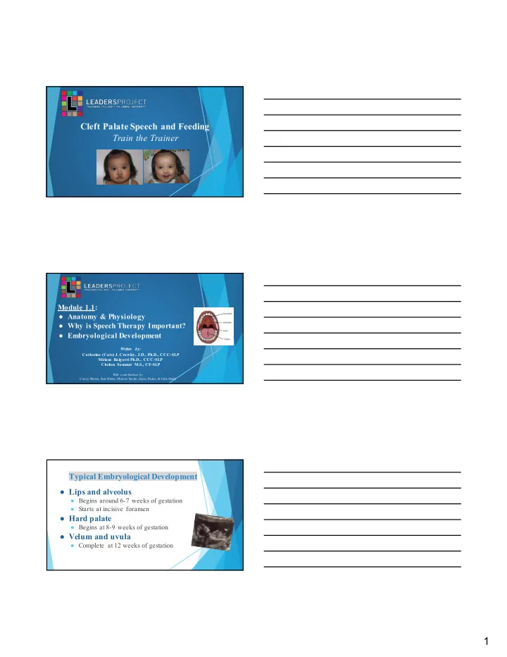

Cleft Palate Speech and Feeding Train the Trainer Module 1.1: ● Anatomy & Physiology ● Why is Speech Therapy Important? ● Embryological Development Written by: Catherine (Cate) J. Crowley, J.D., Ph.D., CCC-SLP Miriam Baigorri Ph.D., CCC-SLP Chelsea Sommer M.S., CF-SLP With contributions by: Casey Sheren, Sara Horne, Marcos Sastre, Grace Frutos, & Julie Smith Typical Embryological Development ● Lips and alveolus ● Begins around 6-7 weeks of gestation ● Starts at incisive foramen ● Hard palate ● Begins at 8-9 weeks of gestation ● Velum and uvula ● Complete at 12 weeks of gestation 1
What does typical anatomy of the oral structures look like? Tensor veli palatini Tensor veli palatini Levator veli palatini An illustration of the muscles involved in velopharyngeal closure What does typical anatomy of the oral structures look like? 2
What does typical anatomy of the oral structures look like? How do the oral structures develop? The incisive foramen is a point of embryological development. From this location the premaxilla closes on the An analogy for the right side and left side forward to the development of a cle ft lip. The palate then closes from the incisive foramen back to the uvula. When one point of development does not close, this results in the cleft. An analogy for typic a l development Typical hard and soft palate 3
Your turn! Turn to your partner and, with a flashlight, examine his/her oral structures. Check the color of the oral tissues, and be sure to identify the: ● Hard palate ● Soft palate ● Uvula Module 1.2: ● Anatomy & Physiology of Different Types of Clefts Written by: Catherine (Cate) J. Crowley, J.D., Ph.D., CCC-SLP Miriam Baigorri Ph.D., CCC-SLP Chelsea Sommer M.S., CF-SLP With contributions by: Casey Sheren, Sara Horne, Marcos Sastre, Grace Frutos, & Julie Smith Unilateral Cleft Lip Mugisha, a child with a unilateral cleft lip from Rwanda. 4
This photo show s that the lip did not finish closing, resulting in a right complete unilateral cleft of the lip. It is complete because it extends into the nostril/nares. Bilateral Cleft Lip Andrea, a child with a bilateral cleft lip. Before and after surgery. Here we see that both sides did not close, resulting in this bilater a l complete cleft of the lip. 5
One can have both a bilateral complete cleft of the lip and a cle f t of the palate as w ell, meaning that during embryological development no closure oc curred. What does typical anatomy of the oral structures look like? Tensor veli palatini Tensor veli palatini Levator veli palatini Here we see that the premaxilla is protruded, which typically contains te e th buds. Premaxilla 6
The bulging premaxilla results from incomplete closur e of the seams anterior to the incisive foramen. If the seams had Premaxilla closed during development, the premaxilla would be correctly placed. An analogy for the development of a cleft Types of Cleft Lip Deformities Clinical Questions ❖ Unilateral (one side) ❖ Bilateral (two sides) Ask yourself: Is one side affected, or both? (Unilateral or bilateral) ❖ Complete (cleft to the nose) Ask yourself: Does the ❖ Incomplete (Only a cleft of cleft go up to the nose? the lip. The nose is not (Complete or incomplete ) impacted) Typical Facial Anatomy 7
Unilateral Incomplete Cleft Lip Unilateral Complete Cleft Lip Bilateral Complete Cleft Lip 8
Cleft Palate Classification We will discuss this later! Turn to your partner and discuss: Your turn! What happened during embryological development that w ould result in this kind of a cleft? ANSWER This is a cleft of the hard palate. It formed during embryological development due to an interruption to closure of the palate from the incisive foramen back to the uvula. 9
Anatomical Variations in Cleft Palate We can see a cleft of just the (& left unilateral cleft lip) soft palate (left) or a cleft of the hard and soft pa late (right) , depending on the point at which development is interrupted. Your turn! Describe the type of cleft you see in the following photos and think about why this might have occurred during development. 10
Answer: Cleft of the hard and soft palate Answer: Bilateral cleft of the lip with bulging premaxilla 11
Answer: Left unilateral complete cleft of the lip 12
Answer: Unilateral complete cleft lip w ith a bulging premaxilla and erupted tooth Module 1.3: ● Submucous and Occult Clefts Written by: Catherine (Cate) J. Crowley, J.D., Ph.D., CCC-SLP Miriam Baigorri Ph.D., CCC-SLP Chelsea Sommer M.S., CF-SLP With contributions by: Casey Sheren, Sara Horne, Marcos Sastre, Grace Frutos, & Julie Smith Three characteristics of a submucous cleft ● Bifid uvula ● Zona pellucida ● Notch in posterior border of the hard palate 13
Submucous Cleft ● Zona pellucida ● Bluish area in the middle of the velum. ○ Bluish coloring ○ Caused by thin mucosa ○ Lack of normal underlying muscle mass ● Velum may appe ar to be in an inverted “V”, especially during phonation. ○ “V” shape ○ Abnormal insertion of the veli pa latini muscles in the posterior section of the hard palate ○ With phonation, velum appears to “tent up” toward hard palate. Submucous Cleft - Zona pellucida Submucous Cleft - “Inverted V” 14
Submucous Cleft - “Inverted V” Submucous Cleft -- Bifid Uvula ❖ May be split down the middle with two pendulous structures ❖ May appear as one structure with line down the center ❖ May have a simple indentation at the posterior border ❖ Uvula may appear small and undeveloped-- hypoplastic. Submucous Cleft -- Bifid Uvula In this photo, we see that there is a submucous cleft with a bifid uvula, as this did not close in development. Submucous cleft is not always identified because patients are not always symptomatic and, even with physical signs of submucous cleft, can have normal speech! 15
Submucous Cleft -- Bifid Uvula Submucous Cleft -- Notch in Posterior Border of Hard Palate ● In normal palate, can often feel slight projection of posterior nasal spine. ● If there is an appreciable notch in the posterior border of the hard palate, this indicates the presence of a submucous cleft palate. ● Use gloved examination. Notch can be small and narrow so use pinky finger to feel. Occult Cleft ● Sometimes children may seem hypernasal, however, there is no physical abnormality in the palate. ● Occult cleft are diagnosed through nasoendoscopy, which is when a scope with a camera is passed through the nostrils to observe how velopharyngeal structures move during speech. 16
Module 1.4: ● Velopharyngeal Closure Written by: Catherine (Cate) J. Crowley, J.D., Ph.D., CCC-SLP Miriam Baigorri Ph.D., CCC-SLP Chelsea Sommer M.S., CF-SLP With contributions by: Casey Sheren, Sara Horne, Marcos Sastre, Grace Frutos, & Julie Smith Muscles Involved in Velopharyngeal Closure ● Levator veli palatini- main muscle for velar elevation ● Superior pharyngeal constrictor- medial displacement of lateral pharyngeal walls ● Musculus uvulae- contracts during phonation and create bulge on velum which adds stiffness of velum ● Palatoglossus muscles- depresses the velum *Tensor veli palatini- opens the Eustachian tube for m iddle ear drainage, contributes little or nothing with velopharyngeal closure. 17
Remember what typical anatomy of the oral structures looks like: Tensor veli palatini Levator Veli Palatini The levator veli palatini muscle cannot connect where there is a cleft palate, meaning that the soft palate cannot raise appropriately to create high pressure oral sounds. The Door Metaphor is an analogy for better understanding cleft palate and why speech errors occur. Play Video #1 entitled “Door Metaphor for Velopharyngeal Closure” 18
Your turn! Turn to your partner and practice reciting The Door Metaphor. This will be necessary when explaining cleft palate airflow and speech to parents of children with cleft palate. Velopharyngeal Closure Patterns There are 4 typical ways velopharyngeal closure can occur. These are different ways in which “the door” can close to create high pressure oral sounds, such as “p”, “b”, “t”, “d”, “k”, “g”, “f”, “s”, “z”, “ch”, “sh”, etc. PPW = Posterior pharyngeal wall “Passav an t’s RLW = Right lateral pharyngeal wall With sag ittal With circu lar With co ro n al Rid g e” is a clo su re p attern , clo su re p attern , LLW = Left lateral pharyngeal wall clo su re p attern , b u lg e o f tissu e mo v emen t o f th e mo v emen t o f th e su p erio r o n th e p o sterio r SP = Soft palate lateral p h ary n g eal lateral p h ary n g eal mo v emen t o f th e p h ary n g eal wall walls is th e main walls an d SP so ft p alate is th e VPC = velopharyngeal closure th at aid s in VPC co n trib u to r to VPC co n trib u te eq u ally main co n trib u to r to VPC to VPC What is velopharyngeal dysfunction (VPD)? Condition where the door--the velopharyngeal closure-- does not happen. Why? Structural - “VP insufficiency” ● Velum too short to reach the posterior pharyngeal wall ● Hole in the palate--a cleft palate--that is a structural reason why the door cannot close Functional - “VP incompetency” ● Physiological: The levator veli palatini does not do its job of lifting the soft palate ● Neurological: Apraxia, dysarthria, brainstem tumor 19
Recommend
More recommend