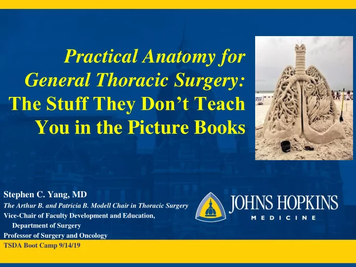

Practical Anatomy for General Thoracic Surgery: The Stuff They Don’t Teach You in the Picture Books Stephen C. Yang, MD The Arthur B. and Patricia B. Modell Chair in Thoracic Surgery Vice-Chair of Faculty Development and Education, Department of Surgery Professor of Surgery and Oncology TSDA Boot Camp 9/14/19
Disclosures No financial disclosures Modest experience, don’t claim to know everything Conflict: I’m a Dukie
Objectives Review important anatomic landmarks in general thoracic surgery Recognize the common anatomic anomalies encountered during these procedures Describe the operative implications of these anomalies
Bronchoscopy Know your scope!
Bronchoscopy Know your scope! Tracheal RUL bronchus
Bronchoscopy Know your scope! Tracheal RUL bronchus Sup seg take off varies
Bronchoscopy Know your scope! Tracheal RUL bronchus Carina Sup seg take off varies Troubleshooting malpositioned double RUL lumen tubes BI Prox R Main
Bronchoscopy – Segmental Nomenclature (anatomic vs Boyden’s)
Mediastinoscopy 1 st PA Main PA BRANCH
Sternotomy, tracheostomy High riding innominate artery
• What is seen here? Azygous lobe
1891 – Tuffier, first successful lung resection for TB 1908: Babcock, RLL lobectomy 1931: Churchill, dissection lobectomy 1933: Graham, left pneumonectomy for lung cancer
Lung Resections 3D vascular anatomy difficult via VATS (thus appreciate open experience) Anatomic anomalies are frequent Increasing number of (VATS) segmentectomies given screening programs picking up small lesions
Nodule Localization Increased incidence w CT screening Use 3-D recon Landmarks: Xiphoid Table position Sup seg tip IPV Nipples
LLL
Pulmonary Collaterals: Pores of Kohn Interalveolar connections, Canals of Lambert Account for: Ventilation across segments and fissures Failure of endobronchial valves Local recurrence after wedge resection
Common PA Variants - Right Lobe Common Variant RUL Truncus anterior 15% no post asc Post asc branch 5% post asc from sup seg RML 55% one common trunk 5% > 2 branches 45% two branches RLL 5 distinct branches or 20% have Post Seg common trunk to basilar multiple sup seg RUL Sup Seg
Common PA Variants - Right Lobe Common Variant RUL Truncus anterior 15% no post asc Post asc branch 5% post asc from sup seg RML 55% one common trunk 5% > 2 branches 45% two branches RLL 5 distinct branches or 20% have common trunk to basilar multiple sup seg
Common PA Variants - Right Lobe Common Variant RUL Truncus anterior 15% no post asc Post asc branch 5% post asc from sup seg RML 55% one common trunk 5% > 2 branches 45% two branches RLL 5 distinct branches or 20% have common trunk to basilar multiple sup seg
Common PA Variants - Left Lobe Common Variant LUL Random order of seg 10% lingular branches branches: none 2-8 may arise or arise proximally Anomalous Sup Seg Lingular LLL 70% sup seg branches off 30% < 2 PA PA before lingula branches to sup LUL 60% single common seg Desc PA bronchus basilar trunk SPV
Common PA Variants - Left Lobe Common Variant LUL Random order of seg 10% lingular branches branches: none 2-7 may arise or arise proximally LLL 70% sup seg branches off 30% < 2 before lingula branches to sup 60% single common seg basilar trunk
Common PV Trunk L>R Reported 14% cases Identify both SPV SPV IPV and IPV If accidentally divided, convert to open, reanastomose to LA (not completion pneumonectomy)
Inferior Pulmonary Ligament Station 9 LN Vascularity increases with inflammation (esp cystic fibrosis) Pulmonary sequestration systemic arterial supply Chyle leak
Operative Pitfalls During VATS Lung Resections RUL: ligate RML PV, injury to PA during dissection behind RUL PV, azygous v. injury, dividing R mainstem bronchus RML: avulsion med seg branch RLL: dividing RML bronchus when completing lower oblique fissure, damage phrenic nerve LUL/LLL: multiple PA branches, dividing L mainstem bronchus, single PV
Intercostal Muscle Flap Do not wrap Take down 1 st after circumferentially! opening ICS
Tissue Flaps of the Chest
Lymph Node Dissection/Sampling
VATS Ports Scapular Tip
Thoracic Duct Injuries: nodal dissection, esophageal mobilization 20% with anomalous anatomy Some advocate ligation during thoracic portion
Esophagus 4 points of narrowing Watch for aberrant or replaced L hepatic a. (25%) Upper path: R chest Lower path: L chest Replaced subclavian – special approaches
Esophageal Dissection
Esophageal Dissection Esophagus R Mainstem
Esophageal Dissection Esophagus Subcarinal LN packet Trachea R Mainstem Divided Azygous
Conclusion A number of common anomalies exist particularly for pulmonary resections Value open operations to aid in VATS/robotics approach Vary operative procedure to gain confidence in anatomy Study CT 3D reconstructions carefully
Thank you syang@jhmi.edu
Recommend
More recommend