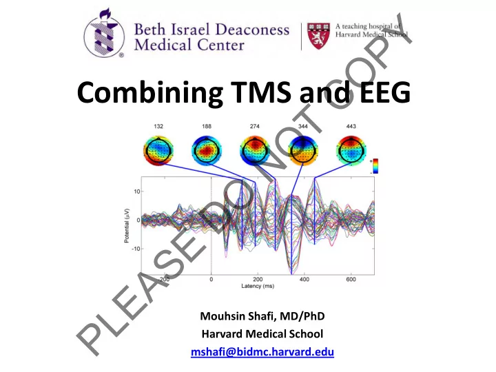

Y P O C Combining TMS and EEG T O N O D E S A E L Mouhsin Shafi, MD/PhD P Harvard Medical School mshafi@bidmc.harvard.edu
Y Talk Overview P O • Intro to TMS and EEG C • Technical issues and challenges T O • Neuroscience Applications of TMS-EEG N – Understanding mechanisms and effects of TMS O – Neurobiology and Cognitive Neuroscience D • Clinical Applications of TMS-EEG E S – Diagnosis A – Monitoring E L – Targeting P
Y TMS: What do we know? P O TMS Protocols C • Single Pulse TMS T • Cortical Mapping O • Motor Threshold N • Central Conduction Time O • Paired Pulse TMS D • One Region • Two Regions E Outcome Measures • Repetitive TMS S MEP Amplitude A • CLINICAL APPLICATIONS E • Across a wide spectrum of L neurologic and psychiatric P diseases
Y This is cool, But … P O What Is Missing? C T O Cortical origin? N Non-motor regions? O State-Dependency? D E Changing brain Motor Responses S MEPS activity states in A disease conditions? E L P
Y EEG to the rescue? P O C T O N O D E S A E L P
Y P EEG: What are we recording? O C T Mostly captures the synaptic activity at the O surface of the cortex. N O EPSP + IPSP generated by synchronous activity of D neurons. E S A Interplay between excitatory pyramidal neurons and E inhibitory interneurons L P
EEG language? Y P O C Amplitude (or Power) Strength T (µ V or µ V 2 ) O N 10Hz Frequency O # of Cycles/Second D (Hz) 20Hz E S A 0 Phase π E (Radians) L P
Y When/How to Record EEG? P O C Continuous Recording (No Event) Event/Stimulus • Anesthesia, T • Sleep O • Resting (eyes open/closed) Trial 1 N O Trial 2 Relative to An Event/Stimulation D • Sensory, motor, cognitive processing E • Electrical stimulation S Trial 100 A E L P Time: Event Related Potential or Evoked potentials Frequency: Event Related Spectral Perturbation Phase
Y How to Analyze EEG? P Time vs. Frequency Domain O C T O N O D Frequency Domain X i ( f ) E imag S Phase A real E L P
Y How to Analyze EEG? P 2 1 O 3 C Local Response T O N - Amplitude/Power O Functional Connectivity - Frequency D Correlation (time) - Phase Coherence (frequency) Synchrony (phase-locking) E Spontaneous EEG: S Θ Spectral Power A Cross-Frequency Phase-Amplitude Coupling EEG + Event: E Event-Related Potentials ( ERP or EP ) L Event-Related Spectral Perturbation P ( ERSP ) Direction of Information Flow Directed Transfer Function Event-Related Synchronization ( ERS ) 1 2 3 Directed Partial Coherence Event-Related Desyncronization ( ERD )
Y In summary what can EEG tell us? P O 1 – EEG is a summation of excitatory and inhibitory C synaptic activity. T 2 – EEG has different spatial, spectral and temporal O architecture under anesthesia, during sleep, in N resting wakefulness, or during sensory processing or higher order cognitive performance. O D Excitability of cortical tissue, and the balance of excitation and inhibition E S Brain state and the integrity of different networks A E Dynamics of interactions within and between L P different brain regions
Y Talk Overview P O • Intro to TMS and EEG C • Technical issues and challenges T O • Neuroscience Applications of TMS-EEG N – Understanding mechanisms and effects of TMS O – Neurobiology and Cognitive Neuroscience D • Clinical Applications of TMS-EEG E S – Diagnosis A – Monitoring E L – Targeting P M/F
Y P O C T O N Marrying TMS with EEG … O the problems … D E S A E L P M/F
Y Initial Problems? P O EEG Amplifiers Saturated! C T O N O D E Ives et al., 2006, Clinical Neurophysiology S A TMS pulse generated too high a voltage (> 50mV) for most E amplifiers to handle. Amplifiers were saturated or even damaged! L P
Y Problem 1 : EEG Amplifier Saturation P O Some Solutions C • De-coupling: TMS pulse is short (.2 to .6ms), so block the amplifier and T reduce the gain for -50µs to 2.5 ms relative to TMS pulse. O Nexstim (Helsinki, Finland) Virtanen et al., Med Biol Eng Comput, 1999; N • Increased Sensitivity & Operational Range: Adjust the sensitivity (100 O nV/bit) and operational range of EEG amplifiers so that amplifiers would not BrainProducts (Munich, Germany) saturate by large TMS voltage D E • DC-Coupling/High Sampling Rate: A combination of DC-coupling, fast 24-bit S analog digital converter (ADC) resolution (i.e., 24 nV/bit) compared to older 16-bit ADC resolution that was limited to 6.1 mV/bit, and high sampling rate (20 kHz)=> capture the A full shape of artifact and prevent amplifier clipping. NeuroScan ( Compumedics ) E L • Limited Slew Rate : Limiting the slew rate (the rate of change of voltage) to P avoid amplifier saturation; Artifact removed by finding the difference between two conditions. Thut et al., 2003; Ives et al., 2006; References: Vaniero et al, 2009; Ilmoniemi et al, 2010
Y P TMS Heated Up O Electrodes! C T O N O One of the subjects had a burn on the skin, to test whether this had anything to do with D rTMS, they placed electrodes on their arm E and stimulated the electrode with different number of stimuli, different intensity and S different duration of stimulation. A E Reference: Pascual-Leone et al., 1990, Lancet L P
Y Problem 2 : Electrode Heating P O Some Solutions C T Small Ag/AgCl Pellet Electrodes O N Virtanen et la., 1999 O D Temp ~ r 2 Temp ~ B 2 E Temp ~ metal electrical S conductivity ( σ ) A E L P
Y There were all kinds of other issues too … P O C • We learned that TMS induces a secondary current (eddy current) in near by conductors. Well… EEG electrodes are T conductors! O High frequency noise in the electrode under the coil N • Movement of electrodes by TMS coil, muscle movement or O electromagnetic force. D Slow frequency movement & motion artifact in EEG E recording S A • Capacitor recharge also induced artifact in the EEG. E Smaller amplitude TMS artifact sometime after L TMS pulse P References : Vaniero 2009; Ilmoniemi 2010;
Other problems Y P O Some Solutions TMS click is loud! C ~ 100 dB 5 cm of the coil Auditory masking with a frequency T matched to the spectrum of the TMS induces auditory TMS click O evoked potentials N Air & Bone Conducted O D E S A E L P Massimini 2005 Nikouline 1999
Y And some remain TMS may cause P motor responses in problematic… O scalp muscles C Frontalis T O N O Temporalis D E Occipitalis Some Solutions S A Changing the coil angle to stimulate Retrieved From: http://education.yahoo.com/referen E muscles less ce/gray/illustrations/figure?id=378 L EMG artifact removal after recording P Independent Component Analysis
Y Site of stimulation is critical P O C T O N O D E S A E L P Mutanen 2012 M /F
Y Other difficulties P O TMS may induced eye blinks C T F3 O F4 N FZ OZ O EOG1 D EOG2 E S A Some Solutions E EOG Calibration Trial L Delete Contaminated Trials P Independent Component Analysis (ICA)
Y P O C Some Tricks!! T O N Minimize residual artifact online (i.e., during recording) O D Removing artifact offline (i.e., after the fact) E S A E L P M/ F
Y Minimizing recorded artifact online P O Coil Orientation with Respect to the Electrode Wires C T O N O - Large positive depression after the stimulus onset for Base, C45, and CC45 directions, D - Residual artifacts were negligible at both 90 positions E S Solution: Rearrange the lead wires relative to the coil orientation. A E L P Results from: H. Sekiguchi et al., Clinical Neurophysiology
Y Minimizing recorded artifact Offline P O Deleting, Ignoring, or ‘Zero-Padding’ C Remove by setting the artifact to zero References: Esser 2006; Van Der Werf and Paus 2006; Huber 2008; Farzan 2010; T O Temporal Subtraction Method Create a temporal template of TMS artifact and subtract it; Example: TMS only N condition; TMS+Task Condition, then subtract TMS Only from TMS+Task References: Thut et al. 2003; 2005. O Removing Artifact and Interpolate D Interpolation: Cut the artifact and connect the prestimulus data point to artifact free post stimulus E Refereces: Kahkonen et al. 2001; Fuggetta et al. 2005; Reichenbach et al. 2011. S PCA and ICA Parse out EEG recording into independent (ICA) or principle (PCA) components and remove A the component that are due to noise; E References: Litvak et al. 2007; Korhonen 2011 Hamidi 2010; Maki & Ilmoniemi 2011; L Hernandez-Pavon 2012; Braack 2013, Rogasch 2014 P Filtering Non-linear Kalman filter to account for TMS induced artifact References: Morbidi et al., 2007 M/F
Y ICA can remove artifactual components P O C T O N O D E S A E L P Rogasch et al, NeuroImage 2014: Used ICA to remove components that are likely muscle and decay artifacts related to stimulation
Y Raw **Clean** P O C T O Slow Blink N Decay Significantly Different O from Clean D E S Bad AEP A electrodes E L P Rogasch et al., NeuroImage , 2014
Y P O C T O N O D E S A E L P
Y P O C T Take Home Message O N What do I need to do if I O want to go back home and D try this? E S A E L P M/F
Recommend
More recommend