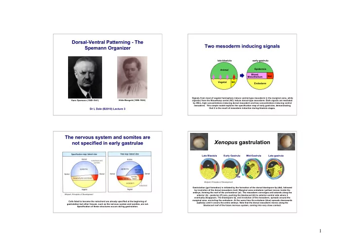

Dorsal-Ventral Patterning - The Two mesoderm inducing signals Spemann Organizer late-blastula early-gastrula Epidermis Animal Blood Not Mesothelium Vegetal NC Endoderm Signals from most of vegetal hemisphere induce ventral-type mesoderm in the marginal zone, while Hilde Mangold (1898-1924) Hans Spemann (1869-1941) signal(s) from the Nieuwkoop centre (NC) induce dorsal-type mesoderm. Both signals are mediated by XNrs, high concentrations inducing dorsal mesoderm and low concentrations inducing ventral mesoderm. This simple model explains the specification map of early gastrulae, demonstrating that it is the result of mesoderm induction during blastula stages. Dr L Dale (B2010) Lecture 3 The nervous system and somites are Xenopus gastrulation not specified in early gastrulae Late Blastula Early Gastrula Mid-Gastrula Late-gastrula A P ar bl P A P A bl dbl Wolpert, Principles of Development Gastrulation (gut formation) is initiated by the formation of the dorsal blastopore lip (dbl), followed by involution of the dorsal mesoderm (red). Marginal zone endoderm (yellow) moves inside the embryo, forming the roof of the archenteron (ar). The mesoderm converges and extends along the Wolpert, Principles of Development anterior (A) - posterior (P) axis, pushing the blastocoel (bl) to anterior-ventral side where it eventually disappears. The blastopore lip, and involution of the mesoderm, spreads around the marginal zone, encircling the endoderm. At the same time the ectoderm (blue) spreads downwards Cells fated to become the notochord are already specified at the beginning of (epiboly) until it covers the entire embryo. Note that the dorsal mesoderm moves along the gastrulation but other tissues, such as the nervous system and somites, are not. blastocoel roof of the future nervous system, coming into very close contact. Specification of these structures occurs during gastrulation. 1
Spemann-Mangold Organizer Graft The nervous system is specified during gastrulation dbl Smith & Slack, JEEM 78: 299-317 (1983) 1º axis vp s Hans Spemann (1919) transplanted pieces of the animal hemisphere at the beginning nt n 1º axis of gastrulation. He used two closely related species of newt that had different levels of pigmentation, allowing him to discriminate between the transplant and host cells. He found that ectodermal transplants always differentiated according to their new location. 2º axis Presumptive epidermis became neural 2º axis plate and presumptive neural plate 2º axis became epidermis. Transplants of the same cells at the end of gastrulation First performed by Spemann (1917) this graft induces a second dorsal axis on the ventral side of the showed that they now retained their fate. embryo. However, Spemann used embryos of the same species and could not distinguish host from graft Presumptive neural plate formed a second tissues. Mangold repeated the experiment (Spemann & Mangold, 1924) using different species of newt, neural plate on the ventral side of the with different levels of pigmentation, as donors and host. She showed that the notochord (n) was always embryo. This experiment showed that the derived from the graft, while the somites (s) and neural tube (nt) were largely derived from the host. This is nervous system is specified during illustrated by this Xenopus experiment, in which the donor embryo was labelled with horseradish gastrulation. Gilbert, Developmental Biology peroxidase (this enzyme produces a dark pigment that allows donor cells to be detected). The transplant has “dorsalized” ventral tissues. Because of its ability to induce a correctly organized dorsal axis, Spemann named the transplant region “The Organizer”. The inductive properties of the dorsal The organizer is the source of dorsalizing signals blastopore lip change during gastrulation early gastrulae complete 2nd axis mid gastrulae partial 2nd axis Spemann suggested that the organizer is the source of dorsalizing signals that induce the nervous system Dorsal lip grafts at the early gastrula stage produce a complete 2nd axis, while grafts from late gastrula (NT) in the dorsal ectoderm and somites, heart, pronephros in the mesoderm. It may also affect fates in stage produce only a partial 2nd axis that lacks a head. During gastrulation the mesoderm population at the dorsal endoderm. This inspired biochemists to try and identify the factors responsible, using neural the dorsal lip changes, with anterior mesoderm being replaced by more posterior mesoderm. Only the induction in newt gastrula stage animal caps as an assay. However, they form neural tissue in response former is capable of inducing a head. Suggests that there may be two organizers, a head organizer and a to stress and even “dead organizers” induced neural tissue. The identity of the organizer factors remained trunk organizer. elusive until molecular techniques became sufficiently powerful to clone their genes. 2
Ventral expression of dorsalizing Genes encoding dorsalizing factors signals induce a second dorsal axis are localized to the Spemann organizer e SEP ov n n Ventral injection ov follistatin noggin chordin of chordin mRNA Sasai et al. Cell 79: 779-790 (1994) A number of genes have now been identified that are only expressed in the organizer of Xenopus gastrulae. Noggin , chordin and follistatin are shown here and all three genes encode secreted proteins. Injection of chordin mRNA into the ventral marginal zone produces embryos that have a second dorsal Follistatin was first cloned in mammals because of a role in the female reproductive cycle and axis. This embryo was stained with an antibody that detects the notochord (n) and otic vesicle (ov), the “accidentally” discovered to be an organizer gene. Noggin was isolated in a screen for mRNAs that could eye (e) is naturally pigmented. We can see a second notochord with a single otic vesicle at the anterior rescue dorsal development in UV-irradiated embryos, while chordin was discovered in a screen for end. The second axis does not possess more anterior regions of the head, thus Chordin only induces a mRNAs specifically localized to the organizer. All three proteins were shown to have dorsalizing activity. partial dorsal axis. Similar results can be obtained with noggin but less so with follistatin . Chordin is required for organizer function Chordin, Noggin, and Follistatin will also Injection of antisense MO Organizer graft neuralize Xenopus animal caps A C control control l r o Epidermis n t gastrula c o C, N or F Epidermis B D - chordin - chordin Neural Tissue Oelgeschlager et al Dev Cell 4: 219-230 (2003) Antisense morpholino oligonucleotides (MO) that specifically block translation of chordin mRNA were injected into Xenopus embryos. Injected embryos develop with head defects (B) but are otherwise normal. Addition of Chordin, Noggin or Follistatin protein to gastrula stage animal caps induces neural tissue. However, when injected embryos are used for an organizer graft a second axis is not induced (D). Thus Noggin is the most potent with Chordin being more potent than Follistatin. Thus all three proteins display Chordin is required for organizer activity. inductive activities characteristic of the organizer. Antisense MOs are oligonucleotides with a modified backbone that is resistant to degradation. MOs bind to their target mRNAs and prevent translation by dislodging the ribosome complex. They are proving to be a powerful tool for disrupting gene function in Xenopus , fish, sea urchins and cell lines. 3
Recommend
More recommend