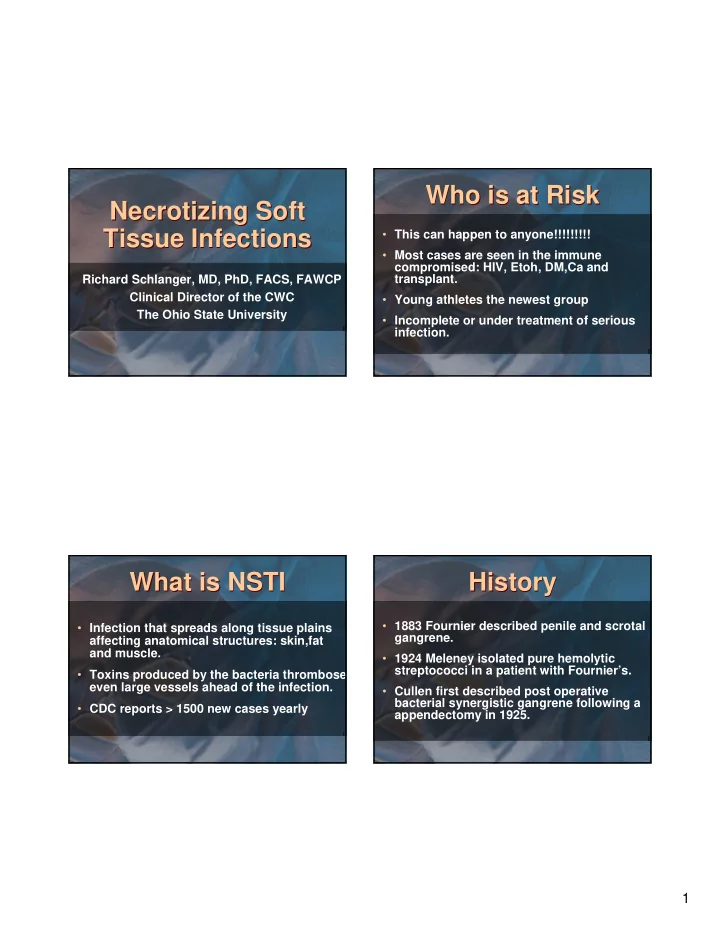

Who is at Risk Who is at Risk Necrotizing Soft Necrotizing Soft Tissue Infections Tissue Infections • This can happen to anyone!!!!!!!!! • Most cases are seen in the immune compromised: HIV, Etoh, DM,Ca and transplant. Richard Schlanger, MD, PhD, FACS, FAWCP Clinical Director of the CWC • Young athletes the newest group The Ohio State University • Incomplete or under treatment of serious infection. What is NSTI What is NSTI History History • 1883 Fournier described penile and scrotal • Infection that spreads along tissue plains gangrene. affecting anatomical structures: skin,fat and muscle. • 1924 Meleney isolated pure hemolytic streptococci in a patient with Fournier’s. • Toxins produced by the bacteria thrombose even large vessels ahead of the infection. • Cullen first described post operative bacterial synergistic gangrene following a • CDC reports > 1500 new cases yearly appendectomy in 1925. 1
History History Etiology Etiology • Necrotizing Fasciitis was first described in • Hypoxia to tissue. 1948. • Decrease in cellular metabolism, defense • Clostridia first cultures in deep tissue reduction of membrane. infection, 1952. • Decrease leukocyte function. • Hemolytic staphylococcus aureus linked to • Localized tissue trauma. the “Flesh Eating Bacteria” 1988. • Reduced host defense. • MRSA the bacteria of the millennium. Classification Classification Diagnosis Diagnosis • HIGH INDEX OF SUSPICION !!!!!!!! • Type of microorganism. • Poor condition of patient. • Type of tissue involved. • Absence of the usual signs of tissue • Therapy required. inflammation. • Rate of progression. • Fever and elevated WBC out of proportion • Initial findings at presentation: bullae, pain to wound. and gas. 2
Diagnostic Aids Diagnostic Aids Necrotizing Fasciitis Necrotizing Fasciitis • Incubation 1- 4 days • Gram Stain of deep tissue. • Onset Acute • Plain radiographs looking for gas or sub-Q air. • Toxicity 2+ • CT scan • Pain Moderate to severe • Culture using deep tissue, not swab. • Exudate Perfuse serosang • Operating Room: debridement • Odor None initially • Gas None initially Necrotizing Fasciitis Necrotizing Fasciitis Clinical Presentations Clinical Presentations • Muscle Viable • Necrotizing Fasciitis • Skin Cellulitic • Necrotizing Cellulitis • Mortality > 35 % • Myonecrosis • Bacteria usually aerobic strep and staph with anaerobic symbiosis +/_ bacteroids and rarely clostridia, e-coli. 3
Necrotizing Cellulitis Necrotizing Cellulitis Myonecrosis Myonecrosis • Incubation 1-3days. • Incubation 1-2 days • Onset Semi- acute • Onset Acute • Toxicity 1+ • Toxicity 3+ • Pain Severe • Pain Out of proportion • Functional loss Acute • Exudate “dirty dish water” • Exudate None • Odor “wet rodent”, foul • Odor None • Gas Present Necrotizing Cellulitis Necrotizing Cellulitis Myonecrosis Myonecrosis • Gas Usually present **** • Muscle Involved: “cooked” • Muscle Dead anatomic groups • Skin Cellulitis and gangrene • Skin Intact • Mortality > 75% • Mortality > 35% • Morbidity > 60% high amp rate 4
The Ohio State The Ohio State Treatment Treatment Experience Experience • The Gold Standard for almost forever was: • 225 patients from 1999- 2008 • Surgery: wide and extensive debridement to viable tissue, reoperation anticipated. • Ages 22 – 82 yo • Antibiotics: broad coverage for aerobic and • > 50% DM anaerobic bacteria • 25% documented immunocompromised • MRSA is public enemy #1 and is presumed • 20% misdiagnosed present and treated. • Mortality depended on time of diagnosis and treatment. Treatment 2010 Treatment 2010 Group 1 Group 1 • Patients transferred to OSU after diagnosis • All major medical and surgical texts: and treatment done at another institution. “Some authors suggest that Hyperbaric Oxygen treatment may have some benefit, • Average delay in transfer 3-5 days but it remains controversial.” Or, “It is • Number of surgical debridement >3 cumbersome and may delay life saving surgery.” • All started on broad spectrum antibiotics • All need redebridement upon arrival at OSU • This is not what we do at OSU !!! • Mortality despite HBO > 75% 5
Group 2 Group 2 Why Hyperbarics Why Hyperbarics • Patients transferred for other facilities • HBO is the placement of a patient in a within 24hrs of diagnosis. closed acrylic tube, pressurized > 1ATA with 100% oxygen. • No treatments done before arrival at OSU • It is bacterialcidal except appropriate antibiotics. • It increases WBC activity • Upon arrival: surgical debridement, HBO, 2 nd look surgery and HBO bid until • It neutralizes toxins symptoms and cultures negative. • It causes angiogenisis. • Mortality < 15% Group 3 Group 3 Surgery Surgery • Role #1 create decompression • All cases seen at OSU and diagnosed fasciotomies in < 12hrs. • Role #2 debride only obviously dead tissue • All cases treated by protocol: antibiotics, • Role#3 2 nd and 3 rd looks after HBO day 1 surgery and HBO (critical care as needed) mandatory • Mortality < 5% • Role #4 amputation is procedure of last resort. 6
7
8
9
Conclusion Conclusion • Always suspect the worst case scenario • Treatment is a three headed monster; massive antibiotics, surgery and HBO • If you do not have direct access to HBO , find the nearest center and get your patient there ASAP. • Mortality is care dependant. 10
Recommend
More recommend