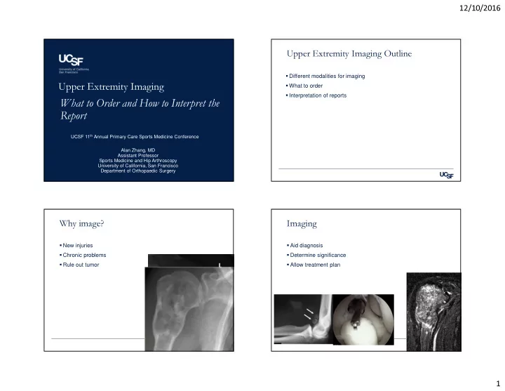

12/10/2016 Upper Extremity Imaging Outline � Different modalities for imaging � What to order Upper Extremity Imaging � Interpretation of reports What to Order and How to Interpret the Report UCSF 11 th Annual Primary Care Sports Medicine Conference Alan Zhang, MD Assistant Professor Sports Medicine and Hip Arthroscopy University of California, San Francisco Department of Orthopaedic Surgery Why image? Imaging � New injuries � Aid diagnosis � Chronic problems � Determine significance � Rule out tumor � Allow treatment plan 1
12/10/2016 Different Modalities Pearls for Ordering Imaging � Write down what you are concerned about � Radiographs • Xrays of wrist with concern of scaphoid fracture � Ultrasound • MRI of shoulder with recurrent instability � CT scan � Radiologists can aid in getting the right studies for you � Bone scan • They can also suggest better studies � MRI Plain radiographs Plain radiographs � Image obtained by projecting of x-ray beams onto a � Good first line evaluation detector � Need multiple views of a joint (AP and lateral) � The amount of ‘whiteness’ is a function of the radiodensity and thickness of the object � Dense object – whiter image 2
12/10/2016 What to order? What to order? � Shoulder � Elbow • AP glenohumeral joint • AP/lateral elbow/oblique joint • Axillary lateral glenohumeral joint What to order? What to order? � Wrist � Forearm • AP/lateral/oblique wrist • AP/lateral forearm 3
12/10/2016 What to order? What to look for? � Hand � Fractures � AP/ Lateral • Displaced • Comminuted • Impacted � Arthritis • Mild, moderate, severe � Abnormal morphology • Spurs, OCD, deformities When to worry? Ultrasound � Uses high-frequency sound waves to produce images � Displaced fractures – always need attention � Similar to sonar wave on getting images of the ocean � Nondisplaced fracture – can immobilize � Stress fracture/ cannot rule out…. � Can be helpful to evaluate ganglion cyst • Need secondary evaluation • Wrist ganglions • Further imaging • Tendon ganglions • Closer follow-up � Diagnose tendon tears • Rotator cuff tears • Rotator cuff repairs 4
12/10/2016 Ultrasound Ultrasound • Can use for targeted therapy � Advantages ‒ Ultrasound guided injections • Non-invasive - Viscosupplementation for Glenohumeral joint • Dynamic - Calcific tendinitis ‒ Tendon instability - Intra-articular injection of the hip � Disadvantage • User-dependent • Cannot image deep tissue • Cannot image tissue within bone CT scan CT scan � Advantages � Tomographic evaluation of the region of interest • Tomographic evaluation � Good for 3D bony anatomy • Gives detail in trabecular and cortical • Glenoid and humeral bone loss structures (better than MRI) � Complex reconstruction ‒ Measure bone loss � Post-traumatic injuries ‒ Evaluate fracture pattern • Wrist malunion ‒ Evaluate healing 5
12/10/2016 Plain Radiographs CT scan � Disadvantages • Subject to metal artifact • Weight limit for obese patients • Higher radiation (1 CT = 229 Xrays) • Contraindicated for pregnant patients 3D CT scan CT Scan � Hamate Fracture 6
12/10/2016 Nuclear imaging Bone scan � Uses radioisotope-labeled biologically active drugs � Rule out tumor – multiple lesions, increase update � Radioactive tracers administered to the patient to serve � Infection – tagged WBC scan as markers of biologic activity � Evaluate symptomatic joints � Images produced by scintigraphy • Such as arthritis • Technetium bone scan • Nonunion • FDG in PET scans • Stress fractures ‒ Measure glycolytic rates ‒ Higher in tumor cells Nuclear medicine MRI � Advantages � Current gold standard for soft tissue injuries • Imaging of metabolic activity ‒ Healed fracture or nonunion • Rotator cuff tears ‒ Arthritis • Labral tears • Diagnosis of infection • Ligament tears � Disadvantages • Cartilage injuries • Lack detail and spatial resolution • Limited early sensitivity � Uses a magnetic field to generate nuclear spin in hydrogen atoms ‒ Fractures usually takes up to several days to show • Relaxation times recorded as radiofrequency signal ‒ Low sensitivity for lytic problems ‒ Multiple myeloma 7
12/10/2016 MRI with contrast -Gadolinum MRI- Gadolinum � Intra-articular contrast � Intravenous contrast • Distends the joint � Evaluate vascularity • Enable evaluation of ligament and labrum • Tumor • Small rotator cuff tears • Post-surgical changes, such as scar tissue • Cartilage injuries, such as TFCC • Concern with kidney insufficiency and complications MRI Rotator Cuff Tears � Helpful to evaluate cuff integrity � Quality of muscle • Fatty infiltration • Retracted tear � Labral pathology Tear 8
12/10/2016 OCD of the elbow TFCC tear � Triangular FibroCartilage Complex Scaphoid fractures MR Arthrogram � For younger patients � Look for partial thickness RCT � Look for delamination � Look for labral pathology 9
12/10/2016 MR Arthrogram- Elbow MR Arthrogram- Wrist � Evaluate ligament tears � Look for communication between compartments � Look for intraarticular pathology, MCL tear loose bodies, OCD Interpretation of MRI Findings MR Shoulder � Asymptomatic individuals • > 60 y.o. – 54% have cuff tear (28% full, 26% partial) • 40 – 60 y.o.– 4% full, 24% partial • 19 – 39 y.o. – 0% full, 4% partial Careful with Interpretations!!! Treat the patient, not the MRI 10
12/10/2016 MRI for Rotator Cuff Tears Interpretation � Rotator cuff tears � Size of tear • Age of patients • How many tendons? • Older patients – common to have partial cuff tears � Retraction ‒ Conservative treatment • How difficult will it be? • Full thickness cuff tears (esp young patient <65) � Fatty Infiltration ‒ Referral for discussion of treatment • How good will the repair be? � Denervation • How well will recovery be? Labral Tears in Shoulder Labral Tears � Anterior labral tears- � Superior labral tear (SLAP) tear • Instability, recurrent dislocations • Pain with overhead activity in young pt (<45) • Treated with PT and surgery if fails conservative treatment � Degenerative labral tears in older pts (>50) are common • Likely incidental finding � Weakness – not correlated with labral tears 11
12/10/2016 AC Joint MRI Elbow � High signal in AC joint � Evaluate ligament tear • Common MR reading � Cartilage injury � AC arthritis • OCD • Clinical diagnosis � Loose bodies • Superficial joint Radiology Reports – love adjectives! MRI Wrist � Fraying vs Partial tear vs Full thickness tear vs Retracted � Ligament tears tear • Scapholunate ligament tears � Cartilage in homogeneity vs fissure vs flap vs unstable flap vs full thickness cartilage loss • Lunotriquetral ligament tears � Tendon degeneration vs tendinosus vs tear � TFCC tears � Bone AVN Clinical Correlation Recommended… • Kienbock’s disease – lunate AVN 12
Recommend
More recommend