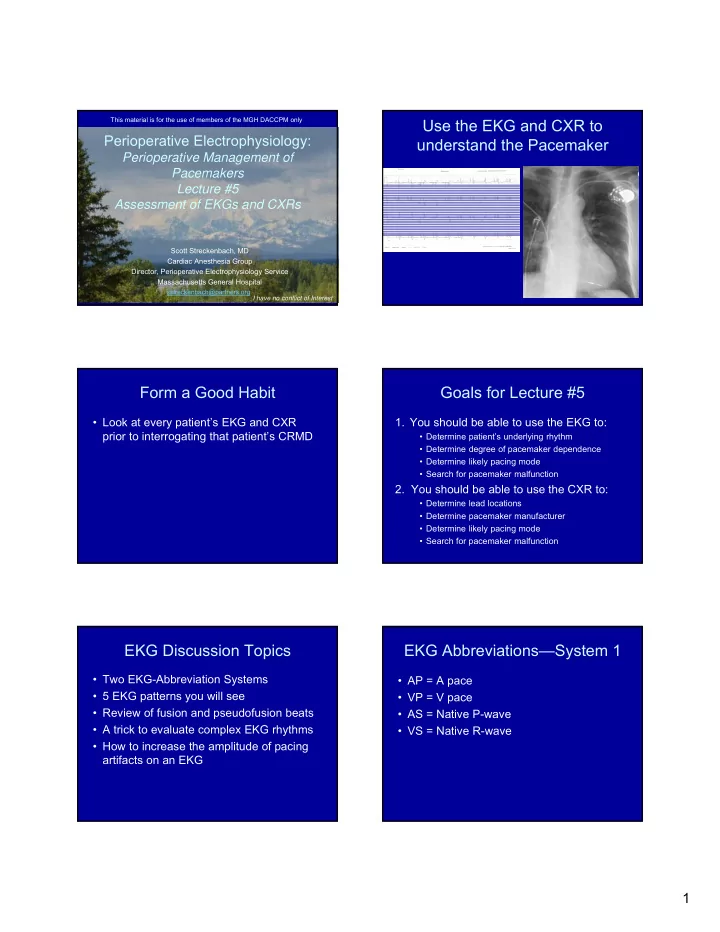

This material is for the use of members of the MGH DACCPM only Use the EKG and CXR to Perioperative Electrophysiology: understand the Pacemaker Perioperative Management of Pacemakers Lecture #5 Assessment of EKGs and CXRs Scott Streckenbach, MD Cardiac Anesthesia Group Director, Perioperative Electrophysiology Service Massachusetts General Hospital sstreckenbach@partners.org I have no conflict of Interest Form a Good Habit Goals for Lecture #5 • Look at every patient’s EKG and CXR 1. You should be able to use the EKG to: prior to interrogating that patient’s CRMD • Determine patient’s underlying rhythm • Determine degree of pacemaker dependence • Determine likely pacing mode • Search for pacemaker malfunction 2. You should be able to use the CXR to: • Determine lead locations • Determine pacemaker manufacturer • Determine likely pacing mode • Search for pacemaker malfunction EKG Discussion Topics EKG Abbreviations—System 1 • Two EKG-Abbreviation Systems • AP = A pace • 5 EKG patterns you will see • VP = V pace • Review of fusion and pseudofusion beats • AS = Native P-wave • A trick to evaluate complex EKG rhythms • VS = Native R-wave • How to increase the amplitude of pacing artifacts on an EKG 1
EKG Abbreviations-System 2 What are the 5 EKG Patterns? • A = A pace Normal Sinus Rhythm • V = V pace A-V sequential pacing (pacer dep) • P = Native P-wave Atrial pacing (SSS) • R = Native R-wave Atrial tracking (AV Block) Ventricular pacing (A Fib) Interpret this EKG How would you describe NSR? • AS-VS • P-R Normal Sinus Rhythm What is the Likely Pacer Setting? Interpret this EKG • DDD • AAI (Sick Sinus Syndrome) • VVI (ICD backup pacing) 2
How would you describe A-V A-V Sequential Pacing Sequential Pacing? • AP-VP • A-V What is the Likely Pacer Setting? Interpret this EKG • Most likely DDD • Could be DOO (magnet applied) How would you Describe Atrial Atrial Pacing Pacing? • AP-VS • A-R 3
What is the Likely Pacer Setting? Interpret this EKG • DDD with long programmed AV interval • AAI How would you Describe Atrial Atrial Tracking (AS-VP) Tracking? • AS-VP • P-V What is the Likely Pacer Setting? Interpret this EKG • DDD • Could be VAT 4
V-Pacing (A Fib) What is the Likely Pacer Setting? • Most likely VVI or VVIR • Could be DDD with VVIR mode switch • Could be DDI or DDIR (least likely) Abbreviations and Patterns Fusion vs Pseudofusion Beats Summary System 1 Description System 2 • We see these often in the OR • Can also see them when analyzing pacers AS-VS Normal Sinus Rhythm P-R on the floor AP-VP A-V sequential pacing (pacer dep) A-V • Recognition of these pacing patterns is AP-VS Atrial pacing (SSS) A-R important in troubleshooting AS-VP Atrial tracking (AV Block) P-V VP Ventricular pacing (A Fib) V • So let’s review Ventricular Fusion Beat Ventricular Fusion Beat Barold SS, Cardiac Pacemakers and Resynch., p. 77 Barold SS, Cardiac Pacemakers and Resynch., p. 77 5
Pseudofusion Beat Pseudofusion Beat Barold SS, Cardiac Pacemakers and Resynch., p. 78 Barold SS, Cardiac Pacemakers and Resynch., p. 78 Baseline NSR Example from the OR with native AV conduction AVI 280 ms • If you have a patient with intact, but A-sense, V-pace prolonged A-V conduction, you can easily AVI 240 ms Non Capture create pseudofusion beats, fusion beats Pseudofusion and finally fully paced beats A-sense, V-pace • Start A-V pacing (DDD mode) with a long AVI 190 A-V interval and progressively SHORTEN Partial capture Fusion beat the pacemaker’s A-V interval A-sense, V-pace PR 140 Full Capture Fusion Beats vs V-Paced Beats Pseudofusion vs Fusion Beats Fusion Beats V-paced Beats Fusion Beat PseudoFusion Beats V-sense We start again with what appears to be a Fusion Beat during A-V pacing Initial assessment reveals narrow paced beats, apparent This time we progressively lengthen the AV interval fusion beats. As the PR interval is shortened, the Notice how the fusion beat becomes a pseudofusion beat (r’ appears, QRS ventricular pacing stimulus captures the entire ventricle and narrows), and then becomes a V-sense beat with pacing inhibition. gives rise to a standard V-paced beat. Note the wider QRS. AV interval started at 180 msec and was increased to 220 msec 6
Key Concept to Remember EKG Discussion Points • Two EKG-Abbreviation Systems • It is nearly impossible to define with certainty a Fusion or Pseudofusion beat • 5 EKG patterns you will see without the presence of a fully paced beat • Review of fusion and pseudofusion beats and a natively conducted ventricular beat • A trick to evaluate complex EKG rhythms • Manipulation of the A-V interval allows one • How to increase the amplitude of pacing to diagnose one beat or the other artifacts on an EKG What can you do if your patient How can you use the with a Pacer has an uncertain programmer to enlarge pacing EKG rhythm? artifacts on the surface EKG? • Interrogate the patient’s pacer with a • Increase the pacing amplitude programmer • Switch the pacing to a unipolar – The atrial and ventricular electrograms will be configuration easier to interpret using the marker channel You need a Sharp Eye to get all Important Message the possible information from the EKG • Always look at the patient’s baseline EKG and the patient’s present rhythm to get at least 2 time points in your evaluation of underlying rhythm 7
Analyze this EKG Analyze this EKG AS-VP AP-VP AS-VP AP-VP AS-VP AP-VP AS-VS Fusion Fusion Analyze this EKG Analyze this EKG Rhythm: AS-VP Patient with a DDDOV pacer What do you notice in V5 CXR Assessment LV-RV 35 msec: LV fires 35 msec before the RV 8
CXR Assessment CXR Basic Anatomy • The CXR is very useful in patients with a pacemaker – How many leads – Pacemaker vs ICD – Manufacturer – Likely pacing mode Ellenbogen, Clin Cardiac Pacing 4 th ed., p.772 The CXR Assessment can be Step by Step CXR Assessment Complicated • Pulse generator – Define the pulse generator location – Confirm the device is a pacemaker – Determine the device manufacturer • Leads – Define lead locations – Are the leads endocardial or epicardial – Are the leads pacing leads or ICD leads – Are the leads connected and positioned correctly? – Are the leads active or passive fixation? Jacob et al, Heart Rhythm Vol 8 No 6 June 2011, p.917 Define Pulse Generator Left Infraclavicular Site Location • More common implantation sites: – Left infraclavicular – Right infraclavicular – Abdomen 9
Right Infraclavicular Site Abdominal Site Ellenbogen, Clin Cardiac Pacing 4 th ed., p.773 Where is the Pulse Generator? Where is the Pulse Generator? S-ICD System Highlights Where is the Pulse Generator? • Single electrode connection • 80 joule (delivered) biphasic shock • Charge time to 80J ≤ 10 seconds • 30 seconds post-shock pacing Boston Scientific 10
Confirm the Device is a CXR Assessment Pacemaker • Define the pulse generator location • Pacers have a radiopaque battery • Confirm the device is a pacemaker • ICDs have a radiopaque battery and capacitor • Determine the device manufacturer • Implantable Loop Recorders are small and usually rectangular • Vagal nerve stimulators typically have a lead going to the IJ vein Pacemaker vs ICD Pacemaker ICD Circuitry Battery Capacitor Pacemakers ICDs Ellenbogen, Clin Cardiac Pacing 4 th ed., p.776, 9 Implantable Loop Recorders CXR Assessment • Define the pulse generator location • Confirm the device is a pacemaker • Determine the device manufacturer Jacob et al, Heart Rhythm Vol 8 No 6 June 2011 11
Two Ways to Determine the Alpha-numeric Code Device Manufacturer • Medtronic M 1. Alphanumeric code S J M 2. Characteristics of the pulse generator • St Jude – Can shape – Battery shape • Bost Sci BOS – “Birth Marks” GDT • Biotronik ET/NT • Sorin ELA Alphanumeric Code What type of Pacer is this? Medtronic Biotronik and Medtronic have characteristic symbols Jacob et al, Heart Rhythm Vol 8 No 6 June 2011, p.918 What type of Pacer is this? Which type of Pacer is this? Ellenbogen, Clin Cardiac Pacing 4 th ed., p.778 12
Boston Scientific Incepta ICD Which type of Pacer is this? Boston Scientific Which type of Pacer is this? What type is this? Biotronik What Type is this? What is the manufacturer? • Alphanumeric code • Characteristics of the pulse generator – Can shape – Battery shape – “Birth Marks ” 13
CXR Algorithm Less than 20% of 1000 pacemakers identified with A-N codes 97% of 2200 pacemakers identified with CaRDIA-X algorithm Jacob et al, Heart Rhythm Vol 8 No 6 June 2011, p.918 My Three Favorite Jacob et al, Heart Rhythm Vol 8 No 6 June 2011, p.918 What Manufacturer? What Manufacturer? 14
What is the Manufacturer? What is the Manufacturer Which Manufacturer? Identify this Device? What Type of Pacer is this? What Type of Pacer is this? 15
Recommend
More recommend