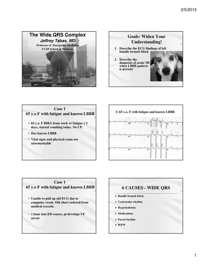

2/5/2013 The Wide QRS Complex Goals: Widen Your Jeffrey Tabas, MD Understanding! Professor of Emergency Medicine 1. Describe the ECG findings of left UCSF School of Medicine bundle branch block 2. Describe the diagnosis of acute MI when LBBB pattern is present Case 1 1) 65 y.o. F with fatigue and known LBBB 65 y.o F with fatigue and known LBBB • 65 y.o. F BIBA from work w/ fatigue x 2 days, started vomiting today. No CP. • Has known LBBB • Vital signs and physical exam are unremarkable Case 1 65 y.o F with fatigue and known LBBB 6 CAUSES - WIDE QRS Bundle branch block • Unable to pull up old ECG due to Ventricular rhythm computer crash. Old chart ordered from medical records. Hyperkalemia Medications • 1 hour into ED course, pt develops VF arrest Paced rhythm WPW 1
2/5/2013 Case 1 - Questions QRS • How do we diagnose LBBB? T Wave P Wave • Can we read ST deviation? Terminal QRS BUNDLE BRANCH BLOCKS The QRS is wide, usually > 0.14 Look at TERMINAL portions of the QRS in Lead V1 and Lead 1 (V6) RBBB = Terminal R in V1 and Slurred S in 1 (V6) LBBB = Terminal R in 1 (V6) and Slurred S in V1 The ST segments are opposite to the terminal portion of the QRS 2
2/5/2013 Left Bundle Branch Man LBBB: Normal ST Segments ACUTE MI in LBBB Iso-electric or Concordant ST segments (same direction Discordant (ST segment opposite the terminal QRS) as QRS) - 1 mm in any lead This is true for every lead OR Excessively Discordant ST segments (opposite QRS but inappropriately large: ST height to S wave height = 1/4 or more) Ischemic Findings in LBBB Ischemic Findings in LBBB Annals of EM, October 2008 • 1 mm Concordant ST elevation – 10 studies with 1,614 patients – Sensitivity = 20% (NLR = 0.8) – Specificity of 98% (PLR = 7.9) • 5 mm Discordant ST elevation – Specificity of 80% (PLR = 4.5) 3
2/5/2013 Ischemic Findings in LBBB Ischemic Findings in LBBB Annals of EM, August 2012 • Excessive Discordance – ST:S wave = 1:4 or more – Sensitivity 58% (vs 30%) ACUTE MI in LBBB ACUTE MI in LBBB EXCESSIVE DISCONCORDANCE CONCORDANT CONCORDANT ST:S wave = 0.25 or more ST Elevation ST Depression 1) 65 y.o F with known LBBB - baseline Another pt with LBBB and Chest Pain c 4
2/5/2013 2) 86 F with CP/SOB and pacer Yet another pt with LBBB and Chest Pain c 86 F with CP/SOB and pacer 2) Prior ECG 1 4 3 5 15 How About LBBB New or Presumed New LBBB “Not Known to be Old?” Current Indications for PCI and Thrombolytics • 1mm ST elevation in 2 contiguous leads or • Left Bundle Branch not known to be old Chang, Am JEM, 2009 • 55 with New LBBB = 7.3% AMI • 136 with Old LBBB = 5.2% AMI • 7746 with no LBBB = 6.1% AMI New LBBB is not predictive of AMI 5
2/5/2013 Take Home Points Neeland, JACC, July 2012 Dx of AMI in LBBB/Pacing • Determine if LBBB is present – LBBB man - Monophasic R in V1, Slurred S in 1 (V6) • Expected ST segments – Opposite terminal QRS – or isoelectric – in all leads For LBBB • Use Sgarbossa criteria • If absent, use rapid serial cardiac biomarkers, bedside echo, or both Take Home Points Pearls Dx of AMI in LBBB/Pacing Dx of AMI in LBBB not known to be old AHA/ACC Approach Acute MI • Presumed New LBBB is an indication for • Concordant ST segments (in same direction as reperfusion QRS) • However, this is not predictive of AMI – 1 mm in any lead My Approach • Excessively Discordant ST segments (opposite • Activate cath lab if moderate suspicion (>25%) direction as QRS) • Initiate thrombolytics for high suspicion (> 50%) – ST:S wave = 1:4 or more • Use cardiac markers or formal echo for the rest. – ST >=5 mm (not as accurate) Involve consultant as early as possible 6
Recommend
More recommend