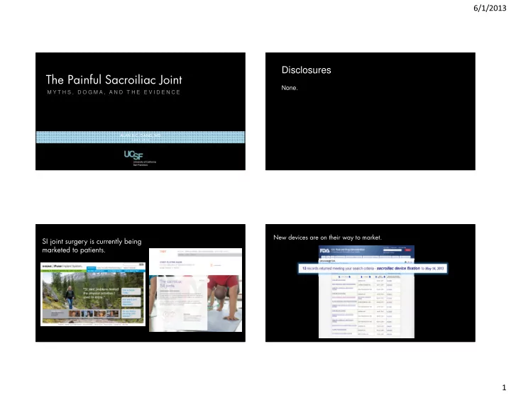

6/1/2013 Disclosures The Painful Sacroiliac Joint None. M Y T H S , D O G M A , A N D T H E E V I D E N C E ALAN B.C. DANG, MD June 1, 2013 New devices are on their way to market. SI joint surgery is currently being marketed to patients. 1
6/1/2013 DOES SI JOINT PAIN COME FROM THE SI JOINT? It’s a diarthrodral joint; all joints can develop arthritis. YES Patients with inflammatory arthritis develop pannus. Patients respond to local anesthetic injections/surgery Articular cartilage is only present on sacral side. NO Precise innervation is still debated. There are no pathognomonic exam findings or radiographic signs for SI joint dysfunction. Anatomy Articular cartilage on sacral surface. Symptomatic Sacroiliac Joints Symptomatic Sacroiliac Joints Fibrocartilage on iliac surface. have abnormal range of motion have abnormal range of motion This mismatch may contribute to degeneration of the joint. Marginal osteophytes can be seen > 50 years old. Incidental MRI changes can be seen > 30 years old. MYTH Radiographic changes can be asymptomatic. Anterior third of the joint has synovial membrane. Posterior portion of the joint is purely ligamentous. 2
6/1/2013 Last year, NASA sent two probes to the moon. Differences in position/velocity from small variations in gravity were used to create a 3-D gravity map. STEREOPHOTOGRAMMETRIC ANALYSIS A pair of X-rays can be used to do the same thing in ROENTGEN STEREOPHOTOGRAMMETRIC ANALYSIS (RSA) No difference in the movement of symptomatic and asymptomatic SI joints. 3
6/1/2013 No difference in the position of the No difference in the position of the SI joints with standing hip flexion test SI joints with manual manipulation. on physical exam. Intra-articular injections & nerve blocks Intra-articular injections & nerve blocks FACT are reliable diagnostic tools for are reliable diagnostic tools for sacroiliac joint dysfunction sacroiliac joint dysfunction There is not a lot of motion at the SI joint MYTH 4
6/1/2013 PROSPECTIVE DOUBLE BLIND STUDY PROSPECTIVE DOUBLE BLIND STUDY L5 Dorsal Ramus + S1-S4 Lateral Branch Block L5 Dorsal Ramus + S1-S4 Lateral Branch Block followed by ligamentous probing/capsular distension followed by ligamentous probing/capsular distension Patient-to-Patient Variability? CONTROL GROUP LIDOCAINE GROUP 100% 60% MAYBE REPORTED PAIN REPORTED PAIN Technical Difficulty of Injections? DEFINITELY ANATOMIC STUDY Fluoroscopically guided S1 and S2 lateral branch blocks with green dye no single finding, or constellation of examination in cadavers followed by dissection findings predicts a positive or negative response to (n = 11) SI joint block from local anesthetic 36% inadequate physical exam vs. inadequate “gold standard” ACCURACY 5
6/1/2013 Is there even any evidence supporting Intra-articular injections & nerve blocks Intra-articular injections & nerve blocks MYTH SI joint dysfunction as a “real diagnosis”? are reliable diagnostic tools for are reliable diagnostic tools for sacroiliac joint dysfunction sacroiliac joint dysfunction Intra-articular injections & peri-articular TRUE blocks provide 6 to 12 months of pain YES relief from sacroiliac joint dysfunction RADIOFREQUENCY ABLATION Multiple studies including randomized, double- Only targets the posterior portion of the joint blind placebo-controlled studies and single-blind (whereas the “degenerating” synovial portion is anterior). placebo-controlled studies show superior pain TRUE relief with steroid injections for SI joint pain vs. placebo. Provides relief of SI joint pain at 3 and 6 months in a formal meta-analysis STUDIES ARE HETEROGENOUS TRUE Mix of CT and Fluoroscopy Aydin SM, Aydin SM, Aydin SM, Aydin SM, Gharibo Gharibo CG, Gharibo Gharibo CG, Mehnert CG, CG, Mehnert Mehnert Mehnert M, and M, and M, and Stitik M, and Stitik Stitik TP Stitik TP TP TP. The role of radiofrequency ablation for sacroiliac joint pain: a meta-analysis. PMR 2: 842-851, 2010. Mix of intra-articular and peri-articular injections 6
6/1/2013 SI joint dysfunction must exist. Superior pain relief with some therapeutic interventions over placebo Early Industry-Funded Studies Literature supports efficacy of Support Surgical Intervention non-surgical therapies. Including a handful of double-blind placebo controlled randomized studies NON-RANDOMIZED INDEPENDENT CHART REVIEW n = 31 NON-RANDOMIZED CASE-SERIES 52% n = 52 COMPLETE PAIN RELIEF 85% 67% would have surgery again when asked 6 months later COMPLETE/EXCELLENT PAIN RELIEF unclear if surgeon-defined or patient-defined 75% 97% had improvement in pain COMPLETE/EXCELLENT/GOOD PAIN RELIEF unclear if surgeon-defined or patient-defined 7
6/1/2013 Inclusion Criteria The Challenge in Pain unresponsive to “prolonged" non-operative treatment and had complete or near complete pain SI Joint Dysfunction relief with CT-guided sacroiliac injection. is Accurate Diagnosis THIGH THRUST TEST 91% sensitivity 66% specificity Positive response to double infiltration The patient is placed in the supine treated as reference standard. position and the examiner flexes and adducts the patient’s hip. Pressure is then applied as an axial load to the femur in order to produce a posterior shear stress on the SI joint IMAGE PROVIDED BY SI-BONE 8
6/1/2013 Distraction Test COMPRESSION TEST Patient in lateral decubitus position. Examiner provides a compressive 69% sensitivity downward portion. 63% specificity FABER (Patrick Test) The patient is placed in the supine Hip flexion, abduction, and external rotation position and the examiner applies pressure to spread the anterior Gaenslen Test superior iliac spines. Patient supine at the edge of examination table with one leg dangled over the side of the table and contralateral leg actively flexed and held close to the chest. Examiner applies a downward force on the extended leg to stress both SI joints. IMAGE PROVIDED BY SI-BONE NONE OF THESE ARE INDEPENDENTLY VALID 85% 3 or more positive provocative tests sensitivity Summary Distraction Test FABER (Patrick Test) 76% Gaenslen Test Thigh Thrust Test Compression Test specificity 9
6/1/2013 SI joint dysfunction is a difficult diagnosis to make due to limitations in diagnostic tools. NSAIDs, non-opiate analgesics should be first-line therapy. Physical therapy is a reasonable option although there are no Thanks. prospective, controlled studies. Steroid injections and RFA have proven benefits, but localization is difficult. Consider CT guidance for non-responsive individuals. The Rosette Nebula Surgical treatment will likely be more prominent in the future. SAN FRANCISCO, CA Randomized trials are currently on-going. February 9, 2013 Canon EOS-60Da / ISO 1600 / 400mm F5.6 L / Celestron CG5-GT 10
Recommend
More recommend