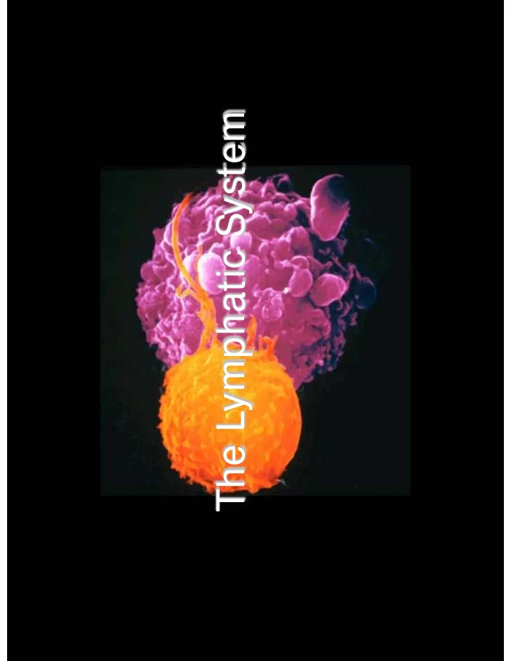

The Lymphatic System
The Lymphatic Systems Overview General Functions Organization Components
Lymphatic System General Functions Transportation Excess fluid from capillary exchange Fats & fat soluble vitamins from lacteals in the small intestines (chyle) Defense B and T cells macrophages Production Lymphocytes within primary lymphatic organs
blood flow Lymph Flow Through F Lymphatic Lymph blood capillary capillaries R Lymphatic collecting System vessels Lymph nodes Venous Return Lymphatic collecting vessels subclavian vein Lymphatic trunks Lymphatic ducts
Lymph Capillaries Located near blood capillaries Differ from capillaries of the cardiovascular system Receive fluid from connective tissue Increased volume of tissue fluid causes one way minivalve flaps open and allow fluid to enter Anchoring filaments stabilize the endothelial flaps High permeability Pros uptake of tissue fluid Cons entrance of bacteria, viruses, and cancer cells Lacteals – specialized lymphatic capillaries Located in the villi of the small intestines Receive digested fats and fat soluble vitamins Fatty lymph – chyle
Location and Structure of Lymph Capillaries
Lymphatic Collecting Vessels Accompany blood vessels Composed of the same three tunics as blood vessels Contain valves Lymph propelled by: Skeletal muscle pump Arterial pulsing Tunica media Lymph node location
Lymph Nodes Cleanse the lymph of pathogens Human body contains approximately 500 lymph nodes Lymph nodes are organized in clusters
Lymph Nodes Function Lymph percolates through lymph sinuses Most antigenic challenges occur in lymph nodes Antigens destroyed – and activate B and T lymphocytes
Lymph Node Structure Major Structures: 1. The lymph node capsule 2. The subcapsular sinus 3. The lymph node cortex beneath the subcortical sinus-the location of primary and secondary lymphoid follicles 4. The paracortex the region surrounding and beneath the germinal centers 5. The medulla deep to the cortex/paracortex, and composed of medullary cords and medullary sinuses 6. Medullary vessels 7. Afferent and efferent lymphatic vessels
Lymph Trunks Formed from the convergence of lymphatic Left jugular Right jugular trunk trunk vessels Right subclavian trunk Left subclavian Five major lymph trunks trunk Right broncho- mediastinal trunk Lumbar trunks Left broncho- mediastinal receives lymph from lower trunk limbs Intestinal trunk receives chyle from Right lumbar digestive organs trunk Bronchomediastinal trunks collects lymph from thoracic viscera Subclavian trunks receive lymph from upper limbs and thoracic wall Jugular trunks drain lymph from the head Left lumbar trunk and neck Intestinal trunk
Lymph Ducts - detail Left jugular Right jugular trunk Thoracic duct trunk Right lymphatic duct Right subclavian trunk ascends along Left subclavian trunk vertebral bodies Right broncho- mediastinal trunk from the cisterna Left broncho- mediastinal trunk chyli Empties into venous circulation at the Right lumbar trunk junction of the left internal jugular and the left subclavian Thoracic duct veins Right lymphatic duct Empties into right internal jugular and Left lumbar trunk subclavian veins Intestinal trunk
Defensive Overview of the Lymphatic System Defenses may be non-specific or specific (immunity) Key cells – lymphocytes & macrophages Recognizes specific foreign molecules Destroys pathogens effectively Also includes lymphoid tissue and lymphoid organs
Specific Defenses (immunity) Types of immunity Cell mediated immunity Antibody mediated (Humoral) immunity Lymphocyte involvement Depends on if B or T cell B lymphocytes – become plasma cells Secrete antibodies Cytotoxic T lymphocytes Destroy antigen-bearing cells
Lymphocyte Development
Lymphoid Tissue Most important tissue of the immune system Primary organs site of development & maturation (bone marrow & thymus) Secondary organs site of activated lymphocyte populations (lymph nodes, lymphatic nodules, spleen) Two general locations Mucous membranes of: Digestive, urinary, respiratory, and reproductive tracts Mucosa-associated lymphoid tissue (MALT or GALT) Lymphoid organs (marrow, thymus, nodes, nodules, spleen)
Thymus Immature lymphocytes develop into T lymphocytes Hassall’s Secretes thymic hormones Corpuscle Most active in childhood Functional tissue atrophies with age Composed of cortex and medulla Medulla contains Hassall’s corpuscles
Spleen Largest lymphoid organ White pulp – thick sleeves of lymphoid tissue Red pulp – surrounds white pulp Composed of: Venous sinuses Splenic cords Two main blood-cleansing functions Removal of blood-borne antigens Removal and destruction of old/defective blood cells Site of hematopoiesis in the fetus (prior to bone marrow development)
Spleen
Tonsils Simplest lymphoid organs Four groups of tonsils Palatine, lingual, pharyngeal, and tubal tonsils Arranged in a ring to gather and remove pathogens Underlying lamina propria consists of MALT
Aggregated Lymphoid Nodules and the Appendix MALT – abundant in walls of intestines Fight invading bacteria Generate a wide variety of memory lymphocytes Aggregated lymphoid nodules (Peyer’s patches) Located in the distal part of the small intestine Appendix - tubular offshoot of the cecum
Disorders of the Lymphatic and Immune Systems Chylothorax – leakage of fatty lymph into the thorax Lymphangitis – inflammation of a lymph vessel Mononucleosis – viral disease caused by Epstein-Barr virus Attacks B lymphocytes Elephantiasis Blockage of lymphatic vessels by filarial worms
Disorders of the Lymphatic and Immune Systems Hodgkin’s disease malignancy of lymph nodes Non-Hodgkin’s lymphoma Uncontrolled multiplication and metastasis of undifferentiated lymphocytes
Recommend
More recommend