The Lymphatic and Immune Systems Chapter 21 The Lymphatic and - PowerPoint PPT Presentation
The Lymphatic and Immune Systems Chapter 21 The Lymphatic and Immune Systems Lymphatic system Main function is to return excess tissue fluid to blood vascular system Lymphatic vessels collect tissue fluid Immune system
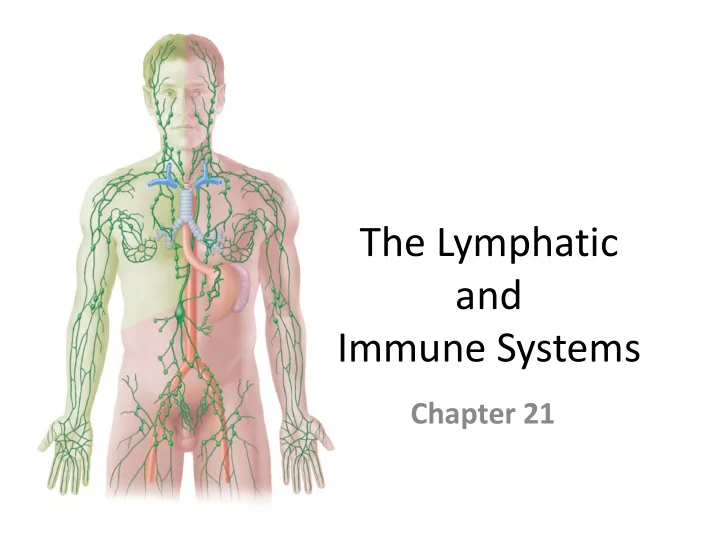
The Lymphatic and Immune Systems Chapter 21
The Lymphatic and Immune Systems • Lymphatic system – Main function is to return excess tissue fluid to blood vascular system – Lymphatic vessels collect tissue fluid • Immune system – Protects our bodies from foreign organisms – Confers immunity to disease – Main components • Lymphocytes, lymphoid tissue, and lymphoid organs
The Lymphatic System • Lymphatic vessels collect tissue fluid from loose connective tissue – Carry fluid to great veins in the neck – Fluid flows only toward the heart – Once tissue fluid is within lymphatic vessels it is termed lymph • Functions of lymphatic vessels – collect excess tissue fluid and blood proteins • Return tissue fluid and blood proteins to bloodstream
Orders of Lymphatic Vessels Venous system Arterial system Heart • Lymph capillaries – s mallest Lymphatic system lymph vessels Lymph duct – First to receive lymph Lymph trunk Lymph node • Lymphatic collecting vessels – Lymphatic collecting c ollect from lymph capillaries vessels, with valves – Lymph nodes are scattered Blood Lymphatic along collection vessels capillaries capillary (a) Structural relationship betw een a capillary bed of the blood vascular system and lymphatic capillaries
Orders of Lymphatic Vessels • Lymph nodes – Scattered along collecting vessels • Lymph trunks – Collect lymph from collecting vessels • Lymph ducts – Empty into veins of the neck
Lymphatic Capillaries • Located near blood capillaries • Receive tissue fluid from CT – Increased volume of tissue fluid • Minivalve flaps open and allow fluid to enter • High permeability allows entrance of – Tissue fluid and protein molecules – Bacteria, viruses, and cancer cells
Lymphatic Capillaries • Lacteals —specialized lymphatic capillaries – Located in the villi of the small intestines • Receive digested fats • Fatty lymph— chyle
Distribution and Features of Lymphatic Capillaries Venous system Arterial system Heart Lymphatic system Lymph duct Lymph trunk Lymph node Lymphatic Tissue collecting fluid vessels, with valves Tissue cell Blood Lymphatic Blood Lymphatic capillaries capillaries capillaries capillary Filaments anchored (a) Structural relationship betw een a capillary to connective tissue bed of the blood vascular system and lymphatic capillaries Endothelial cell Flaplike minivalve Fibroblast in loose connective tissue (b) Lymphatic capillaries are blind-ended tubes in w hich adjacent endothelial cells overlap each other, forming flaplike minivalves. Figure 21.1
Lymphatic Collecting Vessels • Accompany blood vessels • Composed of the same three tunics as blood vessels • Contain more valves than veins do – Helps direct the flow of blood • Lymph propelled by – Skeletal muscles bulging – Nearby arteries pulsing – Tunica media of the lymph vessels • Lymph flow is unaided by heartbeat
Lymph Nodes • Cleanse the lymph of pathogens • Human body contains around 500 • Superficial lymph nodes located in – Cervical, axillary, and inguinal regions • Deep nodes are – Tracheobronchial, aortic, and iliac lymph nodes
General Distribution of Lymphatic Collecting Vessels and Regional Lymph Nodes Regional lymph nodes Internal Cervical jugular vein nodes Entrance of right lymphatic duct into vein Entrance of thoracic duct Axillary into vein nodes Thoracic duct Cisterna Aorta chyli Inguinal Lymphatic nodes collecting Drained by the right vessels lymphatic duct Drained by the thoracic duct Figure 21.2
Microscopic Anatomy of a Lymph Node • Fibrous capsule—surrounds lymph nodes • Trabeculae—connective tissue strands • Lymph vessels – Afferent lymphatic vessels – Efferent lymphatic vessels
Microscopic Anatomy of a Lymph Node Cortex Afferent lymphatic Lymphoid follicle vessels Germinal center Subcapsular sinus Efferent lymphatic vessels Hilum Medulla Medullary cord Medullary sinus Trabeculae Capsule (a) Longitudinal view of the internal structure of a lymph node and associated lymphatics Figure 21.3a
Microscopic Anatomy of a Lymph Node Follicles Trabecula Subcapsular sinus Capsule Medullary cords Medullary sinuses (b) Photomicrograph of part of a lymph node (14X) Figure 21.3b
Microscopic Anatomy of a Lymph Node Macrophage Reticular cells on reticular fibers Lymphocytes Medullary sinus Reticular fiber (c) Reticular tissue w ithin the medullary sinus (540X) Figure 21.3c
Lymph Trunks • Lymphatic collecting vessels converge • Five major lymph trunks – Lumbar trunks • Receives lymph from lower limbs – Intestinal trunk • Receives chyle from digestive organs – Bronchomediastinal trunks • Collects lymph from thoracic viscera
Lymph Trunks • Five major lymph trunks (continued) – Subclavian trunks • Receive lymph from upper limbs and thoracic wall – Jugular trunks • Drain lymph from the head and neck
The Lymphatic Trunks Right jugular trunk Internal jugular veins Esophagus Right lymphatic duct Trachea Right subclavian Left jugular trunk trunk Left subclavian trunk Right subclavian vein Right broncho- Left subclavian vein mediastinal trunk Entrance of thoracic Brachiocephalic veins duct into vein Superior vena cava Left broncho- mediastinal trunk Azygos vein Ribs Thoracic duct Hemiazygos vein Cisterna chyli Right lumbar trunk Left lumbar trunk Inferior vena cava Intestinal trunk (a) Major lymphatic trunks and ducts in relation to veins and surrounding structures, anterior view Figure 21.4a
The Lymphatic Trunks Thoracic duct Aorta Azygos vein on vertebral bodies (b) Thoracic duct (colored green) along the posterior thoracic w all Figure 21.4b
Lymph Ducts • Cisterna chyli – Located at the union of lumbar and intestinal trunks • Thoracic duct – Ascends along vertebral bodies – Empties into venous circulation • Junction of left internal jugular and left subclavian veins • Drains three quarters of the body
Right Lymphatic Duct Right jugular trunk Internal jugular veins Right lymphatic duct • Empties into right Right subclavian trunk internal jugular and Right subclavian vein Right broncho- subclavian veins mediastinal trunk Brachiocephalic veins Superior vena cava Azygos vein Cisterna chyli Right lumbar trunk
The Immune System • Recognizes specific foreign molecules • Destroys pathogens effectively • Key cells— lymphocytes • Also includes lymphoid tissue and lymphoid organs • Lymphoid organs – Lymph nodes, spleen, thymus, tonsils, aggregated lymphoid nodules, and appendix
Lymphocytes • Infectious organisms attacked by inflammatory response – Macrophages, then lymphocytes • Are effective fighters of infectious organisms – Each lymphocyte recognizes a specific foreign molecule • Antigens are any molecules inducing a response from a lymphocyte
Lymphocytes • B lymphocytes and T lymphocytes are the two main classes of lymphocytes • Cytotoxic T lymphocytes – Attack foreign cells directly • Binds to antigen-bearing cells • Perforates cell membrane • Signals cell to undergo apoptosis • Destroy virus infected cells and some cancer cells
Lymphocytes • B lymphocytes – Become plasma cells – Secrete antibodies • Mark cells for destruction by macrophages – Respond primarily to bacteria and bacterial toxins
Lymphocyte Function Target cell, T lymphocyte Dead target cell bearing antigen Antigen 1 T lymphocyte binds to T lymphocyte detaches 2 Target 3 target cell, secretes proteins from target cell. cell dies by that lyse the cell’s membrane, apoptosis. and signals the cell to die. (a) Action of cytotoxic T lymphocyte Antibodies B lymphocyte Plasma cell Bacterium Surface antigen Macrophage B lymphocyte gives rise 1 Antibodies bind to 2 Antibody-coated 3 to plasma cell, which antigens on bacteria, bacteria are avidly secretes antibodies. marking the bacteria for phagocytized. destruction. (b) Differentiation and activity of B lymphocyte Figure 21.5
Lymphocyte Activation • Lymphocytes originate in bone marrow • Some travel to the thymus gland – T lymphocytes • Some stay in bone marrow – B lymphocytes • Able to recognize a unique antigen – Gain immunocompetence • Travels through blood stream – Meets and binds to a specific antigen
Lymphocyte Activation • During activation – Lymphocyte is presented its antigen by • A macrophage • Or a dendritic cell
Lymphocyte Activation • Both T and B lymphocytes produce clones of – Effector lymphocytes • Respond immediately, then die – Memory cells • Wait until the body encounters the antigen again • Basis of acquired immunity • Prevent subsequent infections of the same illness
Recommend


More recommend
Explore More Topics
Stay informed with curated content and fresh updates.