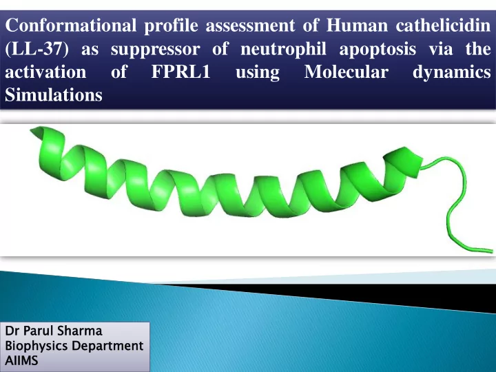

Conformational profile assessment of Human cathelicidin (LL-37) as suppressor of neutrophil apoptosis via the activation of FPRL1 using Molecular dynamics Simulations Dr Dr Parul Sharma Biophysics ics Department ment AIIMS
conserved pro- cationic, frequently peptide α -helical, sequences and a amphipathic N-terminal antimicrobial “cathelin” peptides (AMP). domain. Ca Cathel elicidi icidin prototypes of first-line of defense innovative against infection drugs: infection by serving as broad range of “natural modulate the chemotactic and antibiotics” . immune immunostimulator response. y/ modulatory effects
Apoptosis trend LL LL-37 37 FPRL-1 agoni nist Neutrophil LL LL-37 37 antag agoni nist
Conformational sampling of LL-37 using Structure prediction of FPRL-1 with Molecular dynamics simulations with I-TASSER AMBER force fields 96 and 99SB FPRL1-LL37 potential binding interactions using molecular docking Refinement of docked complexes using molecular dynamics Calculation of ∆G using GBSA and PBSA Preferred binding mode of LL37 depicting molecular interactions with FPRL-1
Available NMR structures of LL37 (in SDS micelle) Wang G., JBC 2008, 283, 32637 (in dodecylphosphocholine Micelles) Porcelli F., Biochem. 2008, 47, 5565
Parameters of MD trjectory ff96 PDB: 2K6O ff99SB
PDB: 2K6O ff96 ff99SB Dark areas are the number of conformations trapped in the configurational space Percent efficiency for generating new patterns in case of MD ff96 was 26% for ff96 and 20%for ff99SB. New patterns have developed earlier (around 5ns) in case of ff99SB as compared to ff96 (after 10ns).
Secondary structural profile of each residue during total simulation time ff99SB ff96 beta turns were again classified into different types using CASICO program type 1 beta turns were found to be maximum, three residue window is considered for a turn in CLASICO Consecutive beta turns resemble 3-10 helix
Secondary structure conformations of LL37 based on H-bonds during MD simulation ff96 ff99SB
Conformations obtained during MD trajectory PDB: 2K6O ff96 ff99SB 10 ns 20 ns 10 ns 20 ns 30 ns 30 ns 40 ns 50 ns 60 ns 50 ns 60 ns 40 ns 70 ns 90 ns 90 ns 70 ns 80 ns 80 ns 100 ns 100 0 ns
Major structural motifs found during MD simulations ff96 ff99 π helix towards C- 3 10 helix from α - helical region is more terminal from ASP 4 – SER 9 . residue ASP 26 - prominent in Arg23-Leu31 ASN 30 70 ns 90ns 100ns α -helix from ASP 4 - LYS 8 towards N- terminal
Final conformations of LL-37 used for docking study with FPRL1 2K6O ff96 ff99
3D Model of Formyl Peptide Receptor like -1 RANK PDB Hit TM-SCORE RMSD IDEN COV. 1 4mbsa 0.821 0.83 0.264 0.832 2 4yay 0.755 1.06 0.268 0.858 3 3oduA 0.727 2.24 0.279 0.792 4 4ea3B 0.717 2.97 0.277 0.806 5 2ksaA 0.708 3.53 0.207 0.823 6 4ib4A 0.695 3.02 0.179 0.798 7 1gzmB 0.694 3.47 0.179 0.812 8 4djhA 0.690 2.99 0.284 0.786 9 1kpnA 0.687 3.61 0.150 0.809 10 1kadA 0.681 4.06 0.183 0.832 Top View Side View
PROCHECK Ramachandran’s map % of re residue ues Most favoured regions 89.7% Additional allowed 8.7% regions Generously allowed 0.3% regions Disallowed regions 1.3% ERRAT2
Prediction of binding site in FPRL-1 for docking of LL-37 PDB: 4MBS; CCR5 chemokine PDB: 4YAY; Human TM-SITE and S-SITE receptor with marketed HIV Angiotensin receptor with its programs drug maraviroc selective antagonist ZD7155
Snapshots taken during MD simulation for FPRL1 docked with 2K6O 1 ns 1.5 ns 0.5 ns 2.5ns ns 2 ns 3 ns 3.5 ns 4 ns 4.5 ns 5 ns
Snapshots taken during MD simulation for FPRL1 docked with helix-bend-helix conformation sampled with ff99SB 1 ns 1.5 ns 0.5 ns 2 ns 3 ns 2.5ns ns 4 ns 3.5 ns 4.5 ns 5 ns
Snapshots taken during MD simulation for FPRL1 docked with kink at K12 conformation sampled with ff99SB 1.5 ns 0.5 ns 1 ns 2 ns 2.5ns ns 3 ns 3.5 ns 4 ns 4.5 ns 5 ns
MMPBSA binding energy calculations Comp mplex ex --- ---> FPRL1-LL37 LL37 (ff96 96) FPRL1 L1-LL37 LL37(ff (ff99S 9SB) B) FPRL1-2K60 2K60 metho hod GENERALI LIZED ED BORN N (at -104.4 .461 617(K (Kcal cal/m /mol) -120.8 .821 217(K (Kcal cal/m /mol) -76.87 8715 15 (Kcal al/m /mol) 1ns) (at 5ns) -108.2 .289 894(K (Kcal cal/mol) -124.3 .381 812(K (Kcal cal/mol) -81.78 7816 16(Kc (Kcal al/mol) POISSON BOLTZ TZMA MANN NN (at -204.0 .003 037(K (Kcal cal/m /mol) -220.7 .733 338(K (Kcal cal/m /mol) -159.2 .290 902(K (Kcal cal/m /mol) 1ns) (at 5ns) -214.0 .054 543(K (Kcal cal/m /mol) -237.6 .642 425(K (Kcal cal/m /mol) -165.4 .432 324(K (Kcal cal/m /mol) 2K6O 6O FPRL1 L1-LL37 LL37(ff99SB) (ff99SB) FPRL1 L1-LL37 LL37 (ff96) 6)
The results demonstrate that the LL-37 has a tendency to attain folded and unfolded conformations, probably due to the low energy barrier between the states accounting for the high level of flexibility of the peptide. Analysis of the MD trajectories of LL-37 depicts the propensity of this peptide to attain α -helices and β -turns under the influence of force fields AMBER ff96 and AMBER ff99SB. With AMBER ff96, conformation of LL37 changed from linear helix to two small helices with kink as proposed by NMR structure (Porcelli, 2008) With AMBER ff99SB force field the peptide attains a stable helical structure with the N and C-terminals being extended and flexible. Docked conformations of peptide by MD using ff99SB with FPRL1 was having maximum interactions and free binding energy as compared to NMR structure. This could further give new insight into the neutrophil apoptosis role of LL-37 Additionaly, the functional diversity of cathelicidin makes it a relevant and lucrative target to investigate.
Recommend
More recommend