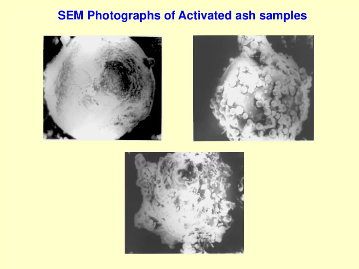

SEM Photographs of Activated ash samples
SEM Micrographs (Original ash samples) (a) Sample S1F1 (b) Sample S2F2 (c) Sample SA35F2 (d) Sample SA50F3 (e) Sample SA35F4 (f) Sample SA50F5
Agglomeration of Ash Particles due to Flue Gas Conditioning (a) Sample CA8S12F1 (b) Sample CA8S12F2 (c) Sample CA8S12F3 (d) Sample CA8S12F4 (e) Sample CA8S12F5 (f) Sample CA8S12F6
SEM micrographs of Silica Fumes & Ground Granulated Blast Furnace Slag (GGBFS/BFS) SF BFS
Determination of fabric structure of fine-grained soils Using SEM Compacted sample Cubic specimen Specimen preparation (Challenges): • Removal of pore fluid from the specimen without disturbing its microstructure. • Freeze-drying technique (for swelling/shrinking type of soils) • Air-drying technique (for non swelling/shrinking type of soils) • Specimen should be able to withstand the vacuum inside the microscope. • As illumination is with electrons, specimen should be made to conduct electricity. • Specimen are coated with a very thin layer of Gold or Carbon (a sputter coater). • Gold coating film can absorb X-ray signal generated into the specimen. • For obtaining X-ray spectrum of a non-conducting sample a coating material very transparent to the X-ray (Carbon) must be utilized.
Face-Face interaction Kaolinite plate stacks
Face-Edge & Edge-Edge interactions
Recommend
More recommend