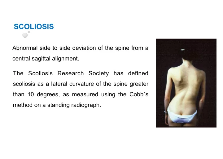

SCOLIOSIS Abnormal side to side deviation of the spine from a central sagittal alignment. The Scoliosis Research Society has defined scoliosis as a lateral curvature of the spine greater than 10 degrees, as measured using the Cobb´s method on a standing radiograph.
• Classification – Causal • Structural (failure to correct). 1. IDIOPATHIC (most common type: 80%). 2. Others: congenital, developmental, neuromuscular, post- traumatic, inflammatory, neoplastic, bone softening • Non structural (functional). – Location: Based On Region of spine in which the apex is located – Direction: – Dextroscoliosis – Convex to the right – Levoscoliosis – Convex to the left 2 2
Idiopathic. 3 types 1. Infantile (<4yo ). Most commonly male Left thoracic curve predominates 75% resolve spontaneously (usually < 30°) 2. Juvenile (4-9 yrs). Female 4:1 Right thoracic curve most common 3. Adolescent (10 yr to skeletal maturity). Female 9:1 Most common type overall and most common type of idiopathic forms Right thoracic curve most common 3 3
Adolescent Idiopathic Scoliosis Red flags: • Severe pain • Left thoracic curve • Abnormal neurologic examination (Specialty consultation and magnetic resonance imaging are needed). Risk factors for curve progression • Large curve magnitude (measured with Cobb´s method). • Skeletal immaturity (assess with Tanner staging and Risser grading). • Female gender 4 4
Risser grade Evaluates growth potential. (0 to 5 based on the degree of bony fusion of the iliac apophysis). 0: no ossification 5: complete bony fusion 5 5
Cobb´s method Using a standard PA standing radiograph of the spine, the Cobb´s angle is formed by a line drawn perpendicular to the top of the superior vertebrae of the scoliotic curve and a similar perpendicular line drawn along the bottom of the inferior vertebrae. The angle between intersecting lines drawn perpendicular to the top of the superior vertebrae and the bottom of the inferior vertebrae is the Cobb angle (here, 62/75 degrees). 6 6
Adolescent idiopathic scoliosis. Prevalence in North America. 2-4% of children between 10 and 16 years of age. From adolescents diagnosed with scoliosis, only 10% have curve progression requiring medical intervention. 7 7
Prognosis Curves <30 degrees at bone maturity are unlikely to progress Curves from 30 to 50 degrees progress an average of 10 to 15 degrees over a lifetime Curves of >50 degrees at maturity progress steadily at a rate of 1 degree per year. In most patients, life-threatening effects on pulmonary function do not occur until the scoliotic curve is 100 degrees or greater. The American Academy of Orthopedic Surgeons recommends screening girls at ages 11 and 13, and screening boys once at age 13 or 14 years of age. 8 8
HISTORY Adolescent idiopathic scoliosis is primarily a diagnosis of exclusion. The history and physical examination are intended to exclude secondary causes for the spinal deformity. 9 9
Physical Examination- Screening. Step 1: Front View Shoulders should be level and at the same height Distance between arm and torso equal on both sides Crest of hips level on horizontal plane Head straight and centered Step 2: Back Standing View Shoulders should be level and the same height Distance between arm and torso equal on both sides Crest of hips level on horizontal plane Head straight and centered Scapula level on both sides 10 10
Step 3: Adam’s Bending Technique The child bends forward at the waist until the spine becomes parallel to the horizontal plane, while holding palms together with arms extended. The examiner looks along the horizontal plane of the spine from the back and side to detect an asymmetry in the contour of the back known as a “rib hump”. A rib hump is a hallmark of scoliotic curves greater than 10 degrees and should prompt radiographic evaluation. 11 11
Step 4 Side View Asses if hyperkhyphosis Step 5 Bending Front View Shoulders level? Is one side of torso more rounded than the other? Look for lumbar prominence 12 12
Step 6 Scoliometer Measurement Before measuring with the Scoliometer, adjust the height of the person´s bending position to the level where the deformity of the spine is most pronounced. Lay the Scoliometer across the deformity at right angles to the body with the 0 mark ever the top of the spinous process. The screening examination is considered positive if the reading on the Scoliometer is 7 degrees or more at any level of the spine. Persons in this category should be referred for further medical evaluation. 13 13
2013 2013 2013 2013 2013 2014 2015 totals Mahalapye Shoshong Shoshong Shoshong Shoshong Shoshong Mahalapye # screened 150 122 229 190 93 200 112 1096 Age 11-14 12-15 10-15 10-15 10-15 8-12 10-15 range # scoliosis 1 0 0 0 0 0 0 1 # female 74 52 92 82 47 100 54 501 # male 76 70 137 108 46 100 57 594 14 14
Action steps for positive findings Students with scoliometer readings over 7-10 degrees or visible deformity refer to WSC or orthopaedic clinic for: • Detailed clinical examination • Consideration for x-rays to measure degree of curvature • Treatment plan: observation, pain management, bracing or surgery 15 15
• Raquel Rojo Delgado • Clinic Supervisor • World Spine Care • www.worldspinecare.org • raquelrojodelgado@gmail.com 16 16
Recommend
More recommend