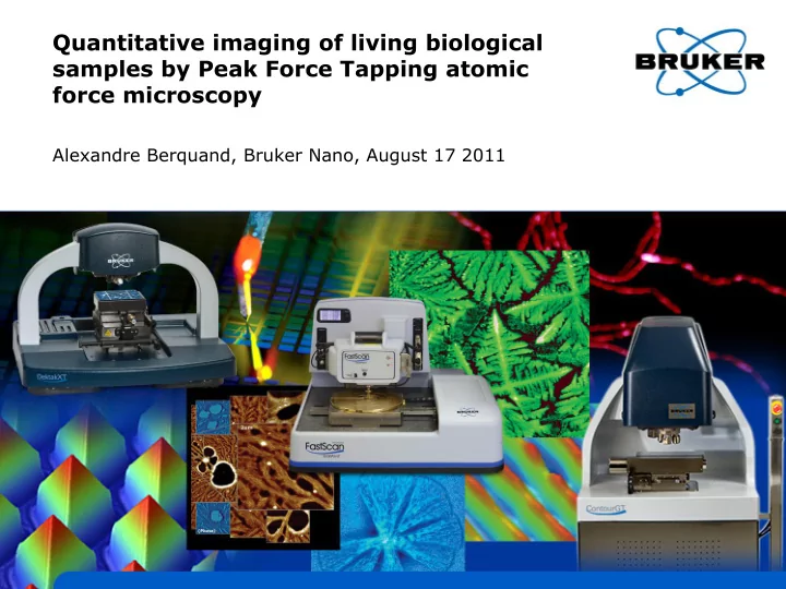

Quantitative imaging of living biological samples by Peak Force Tapping atomic force microscopy Alexandre Berquand, Bruker Nano, August 17 2011
Why force measurements are essential in biology? • Mechanical properties of cells are determined by the dynamic behavior of their cytoskeleton. • Alterations of the mechanical phenotype of the cell can lead to severe malfunctions or disease (cancer, malaria, neurodegeneration). • Cancer cells are known to be softer than their normal homologues. • AFM is the tool of choice to measure cells mechanical properties ex vivo and to correlate a change in mechanical properties with: • Drug treatment • Aging • Pathology 8/17/2011 BRUKER CONFIDENTIAL 2
AFM under physiological conditions • Different types of perfusion systems to keep cells alive for a non-limited period of time: Perfusing Stage Incubator Regular fluid cell
Tapping Mode and Phase imaging depends on AFM parameters, surface and volume properties • The phase shift just reflects the energy dissipated but is a contribution of several factors and is not quantitative 8/17/2011 BRUKER CONFIDENTIAL 4
Force Spectroscopy 2 Stiffness (Young’s force (nN) modulus) 1 0 Adhesion -1 500 0 distance (nm) Single force Force volume • Main drawbacks: slow, poor resolution and lack of information 8/17/2011 BRUKER CONFIDENTIAL 5
Peak Force Tapping - principle • Works with most standard AFM probes in the standard AFM cantilever holders. • Z piezo is driven with sinusoidal waveform (not a triangle as in force-distance curves). • Z drive frequency is 2 kHz (Catalyst 1 kHz). That’s far below the cantilever’s resonance. • Z drive amplitude is fixed at typical value of 150 nm (300 nm peak-to-peak) • Vertical motion of probe produces force- distance plots as it taps on the sample. • Imaging feedback is based on the Peak Force of the force-distance curve. 8/17/2011 BRUKER CONFIDENTIAL 6
Peak Force Tapping - features • SCANASYST: • uses automatic image optimization technology • simplifies and speeds up expert-level image acquisition • PEAKFORCE QNM: • generates quantitative maps of nanoscale material properties • does this simultaneously during imaging at consistently low force and high resolution • Data extraction: 8/17/2011 BRUKER CONFIDENTIAL 7
PeakForce QNM - Calibration • Relative method • Calculate the defl. Sens. • Calculate the spring constant • Image a ref. sample and adjust the tip radius • Adjust the deformation • Absolute method • Calculate the defl. Sens. • Calculate the spring constant • Image a tip check sample and measure the tip radius
PeakForce QNM - Modulus measurement Choose probe type according to range of expected modulus Requirements: Probe needs to deform sample (minimum: a few nm) Probe needs to be deflected by sample (minimum a few nm) 8/17/2011 BRUKER CONFIDENTIAL
Typical example: DNA • PeakForce QNM works in both air and liquid • Relevant and quantitative contrast on all the channels • Applications in liquids have not been as thoroughly explored: • DNA, most of polymers: OK • Cells? 2: Elasticity 3: Adhesion
Any compromise between measurement of mechanical properties and resolution? Simon Scheuring , Physico-Chimie Institut Curie , ScanAsyst lever, 0.4 N/m ) Scale bar 10 nm Scheuring et al Eur Biophys J (2002) 8/17/2011 BRUKER CONFIDENTIAL 11
Sea water samples: imaging of frustules 1 st time that such sample is imaged by AFM • • Very detailed contrast in Young’s modulus and deformation 8/17/2011 BRUKER CONFIDENTIAL 12
Sea water samples: imaging of diatoms • First image of living diatoms with PFT and PFQNM. • YM of different parts: • Fibulae ~200 MPa • Silica stripes ~44 MPa • Core matrix ~21 MPa • … Under press (Journal of Phycology)
Imaging of E. coli K12 • Strain very hard to image by AFM because they move very fast when under stress • b: 3d-height (10x10 m) image of a necklace of living k12 acquired in 20 min. • DMT modulus image of the same bacteria. Average Young’s modulus = 183 kPa 8/17/2011 BRUKER CONFIDENTIAL 14
PFQNM study on human glioblastoma U251-MG cells (invasive) 1 st site-specific 2 nd site-specific recombination: recombination: Integration of expression vector Empty vector + GFP as which carries the gene of interest, integration site inside the GFP site Selection of cells having integrated the vector Test with TP53 and PTEN Possibly have ≠ mechanical properties
PFQNM High Resolution images on glioblastoma - display 2 channels simultaneously 40x40 µm PF error image 3d-height + deformation skin 8/17/2011 BRUKER CONFIDENTIAL 16
PFQNM Low Resolution images on glioblastoma - statistics Topography (z: 0-250 pN) Elasticity (z: 0-1.2 MPa) Adhesion (z: 0-800 pN) • 128x128 images (5 min per image): averaging on a high number of images • Highly quantitative • No damage of the sample Deformation (z: 0-250 nm)
Results & Conclusion Young’s modulus (kPa) Elasticity (kPa) 140 120 100 80 60 40 20 0 TP53 and PTEN induced are Ctrl IND Ctrl non- tp53 non- tp53 IND pTEN non- pTEN IND IND IND IND significantly stiffer and less deformable than the other Deformation (nm) Deformation (nm) cell types 250 200 150 100 50 0 Ctrl IND Ctrl non-IND tp53 non- tp53 IND pTEN non- pTEN IND IND IND
Imaging of living HaCaT and effect of Glyphosate Cell under stress: [Glyphosate] retracting & increase of YM by synthesizing stress factor 3 fibers Adhesion much Average dissipation = 1.3 keV = 2.10 -16 J higher between the cells than on the cells 8/17/2011 BRUKER CONFIDENTIAL 19
MIRO: Overlay optical and AFM data in a few clicks 3) Overlay optical and AFM 1) Import optical image into 2) Target a location for the images Nanoscope AFM scan Hela HaCaT
Combining MIRO and PFQNM • a: overlay of fluorescence (nucleus + actin) and AFM (PF error + YM) images. • b: PF error channel: 0-450 pN • c: YM channel: 0-4 MPa • d: deformation channel: 0-250 nm • Offers nice perspectives in biology: correlate fluorescence and AFM signals simultaneously in response to drug treatment 8/17/2011 BRUKER CONFIDENTIAL 21
Typical samples and corresponding probes - Summary Calibration of Young’s Modulus by Gelatin or Agarose: ~1 to 100 kPa 8/17/2011 BRUKER CONFIDENTIAL 22
Conclusions • Since its development, Peak Force Tapping and PeakForce QNM have greatly improved to extend the range on biological samples • Though it’s still not 100% quantitative for the softest samples, a very wide range of applications can be covered • We are still working on expanding the range… • Promising possibilities for recognition mapping with functionalized probes (still confidential) 8/17/2011 BRUKER CONFIDENTIAL 23
New Application Note released… 8/17/2011 BRUKER CONFIDENTIAL 24
Acknowledgements (sample providers) • Vesna Svetlicic, Tea Radic and Galja Pletikapic (Rudjer Boskovic Institute, Zagreb, Croatia) • Gregory Francius (LCPME, Nancy, France) • Andreas Holloschi, Leslie Ponce, Ina Schaeffer, Hella-Monika Kuhn, Petra Kioshis and Mathias Hafner (University of Applied Sciences, Mannheim, Germany) • Laurence Nicod, Celine Caille and Celine Heu (Institut FEMTO-ST, Besancon, France)
Contact information • alexandre.berquand@bruker-nano.com +49 174 333 94 62 +49 621 842 10 66 • Contact email for Sales and Support ProductInfo@bruker-nano.com • Webinar Series www.bruker-axs.com/atomic-force-microscopy-webinar-series • PeakForce QNM www.bruker-axs.com/PeakForceQNM • ScanAsyst www.bruker-axs.com/ScanAsyst • BioScope Catalyst www.bruker-axs.com/bioscope-catalyst-atomic-force-microscope • Bruker NanoScale World Forum – Share, discuss, and learn about everything nano http://nanoscaleworld.bruker-axs.com/nanoscaleworld/
Recommend
More recommend