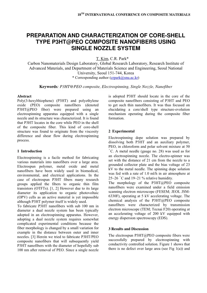

18 TH INTERNATIONAL CONFERENCE ON COMPOSITE MATERIALS PREPARATION AND CHARACTERIZATION OF CORE-SHELL TYPE P3HT@PEO COMPOSITE NANOFIBERS USING SINGLE NOZZLE SYSTEM T. Kim, C.R. Park* Carbon Nanomaterials Design Laboratory, Global Research Laboratory, Research Institute of Advanced Materials, and Department of Materials Science and Engineering, Seoul National University, Seoul 151-744, Korea * Corresponding author (crpark@snu.ac.kr) Keywords : P3HT@PEO composite, Electrospinning, Single Nozzle, Nanofiber is adopted P3HT should locate in the core of the Abstract Poly(3-hexylthiophene) (P3HT) and polyethylene composite nanofibers consisting of P3HT and PEO oxide (PEO) composite nanofibers (denoted to get such thin nanofibers. It was thus focused on P3HT@PEO fiber) were prepared using an elucidating a core/shell type structure-evolution electrospinning apparatus equipped with a single mechanism operating during the composite fiber nozzle and its structure was characterized. It is found formation. that P3HT locates in the core while PEO in the shell of the composite fiber. This kind of core-shell structure was found to originate from the viscosity 2 Experimental difference and shear flow during electrospinning Electrospinning dope solution was prepared by process. dissolving both P3HT and an auxiliary polymer, PEO, in chloroform and polar solvent mixture at 50 ˚ C. A metal needle (gauge no. 28) was used as for 1 Introduction an electrospinning nozzle. The electro-spinner was Electrospinning is a facile method for fabricating set with the distance of 21 cm from the nozzle to a various materials into nanofibers over a large area. grounded collector plate and the bias voltage of 23 Electrospun polymer, metal oxide and metal kV to the metal needle. The spinning dope solution nanofibers have been widely used in biomedical, was fed with a rate of 1.0 ml/h in air atmosphere at environmental, and electrical applications. In the 25~26 ˚ C and 19~21 % relative humidity. case of electrospun P3HT fibers many research The morphology of the P3HT@PEO composite groups applied the fibers to organic thin film nanofibers were examined under a field emission transistors (OTFTs). [1, 2] However due to its large scanning electron microscope (FESEM; JEOL JSM- diameter its application to organic photovoltaic 6330F), operating at 5 kV accelerating voltage. The (OPV) cells as an active material is yet impossible chemical analysis of the P3HT@PEO composite although P3HT polymer itself is widely used. nanofibers were characterized by transmission To fabricate P3HT nanofibers with sub 100 nm in electron microscope (TEM; Tecnai F20) operating at diameter a dual nozzle system has been typically an accelerating voltage of 200 kV equipped with adopted in an electrospinning apparatus. However, energy dispersion spectroscopy (EDS). adopting a dual nozzle system requires somewhat complicated experimental conditions because the fiber morphology is changed by a small variation for 3 Results and Discussion example in the distance between outer and inner The electrospun P3HT@PEO composite fibers were nozzles. [3] Herein we tried to fabricate P3HT/PEO successfully prepared by electrospinning with composite nanofibers that will subsequently yield conductivity controlled solution. Figure 1 shows that P3HT nanofibers with the diameter of hopefully sub there is no defect over large area (see Fig 1(a)) and 100 nm after removal of PEO. Since a single nozzle
the diameter of an individual fiber has the diameter (light region). In this system, PEO solution has high of approximately 100 nm. viscosity. Fig.1. (a) SEM micrographs of homogeneous electrospun P3HT@PEO fibers over large area and (b) a magnified image of (a) with a scale bar of 100 nm. The structure of the composite fibers could be a core-shell type structure or a randomly phase- separated structure. To see which one is the case, element mapping using EDS was carried out for sulfur and oxygen that are typical elements of P3HT and PEO respectively. Fig. 2 illustrates the scanning transmission electron microscopy (STEM) image and mapping image of each element. Due to the low melting temperature (70 ˚ C) of PEO, the STEM and EDS analysis was intentionally performed using very thick fiber. Almost all fibers have the diameter no less than 100 nm. The element mapping image Fig.2. (a) STEM micrograph of electrospun clearly shows that sulfur element locates in the core P3HT@PEO composite fiber and (b) EDS mapping while oxygen at the shell of the composite fibers, with the intensity graph on the selected area of (a). indicating the presence of P3HT in the core and PEO at the shell. This type of composite fiber may be favorable for the preparation of very thin nanofibers of conjugated polymers including P3HT. The core-shell structure formation from a single nozzle system may be unusual. To investigate this core-shell type structure evolution mechanism, the energy dissipation of the fiber was calculated. Energy dissipation rate is given as υ ∝ ∫ d η 2 ( ) (1) E rdr dr where η is the viscosity, ν is the velocity, and r is a radial position from the center in Fig. 3. Basic assumptions of the calculation are similar with a work studied by Williams. [4] To study the energy dissipation rate of the flow, cross section of the flow is divided into three regions. Viscosity of region 2 (dark region) is higher than that of region 1 and 3
PAPER TITLE = ∫ Fig. 3. Schematic image of the flow model. πυ 2 (7) Q rdr Region 1 and 3 have low viscosity η 1, and π K region 2 has high viscosity η 2. = 4 (8-1) Q r 1 1 η 4 1 System of the electrostatic flow is different from π K = 4 − 4 ( ( ) laminar flow due to the surface charge of the flow. Q H r r 2 η 2 1 4 (8-2) Tangential stress at the surface is generated by the 1 surface charge, so the surface velocity (velocity of + − 2 2 − 4 2(1 )( ) H r r r 1 2 1 the region 3) is always larger than the velocity at the π K center (velocity of the region 1). [5] To simplify the = + − 4 − 4 (1 (1 )( ) Q H r r 3 η 2 1 4 calculation, the relative velocity at the center is fixed (8-3) 1 as 0, and the stress profile is assumed to be linear. − − 2 − 2 2(1 )( )) H r r The velocity profiles of each region are calculated 2 1 π K based on integration, = + + = =Q hom o (9) Q Q Q Q 1 2 3 hom η o 4 2 Kr υ = 1 (2-1) 1 η From the equation (8) and (9), 2 1 K ∴ = K υ = 2 + − 2 ( (1 ) ) (2-2) Hr H r K 2 η 1 2 hom o (10) 1 1 K υ = 2 − − 2 − 2 ( (1 )( ) (2-3) r H r r + − − − − − 4 4 2 2 1 (1 )( ) 2(1 )( ) 3 η 2 1 H r r H r r 2 2 1 2 1 1 Therefore, where K is a constant related to the stress profile, H = η 1 / η 2 . According to Eq. (1), the energy dissipation E ∴ = rate can be obtained. E hom o (11) 2 4 K r − − 4 − 4 = 1 (1 )( ) 1 (3-1) H r r E 2 1 1 η 4 + − 4 − 4 − − 2 − 2 2 (1 (1 )( ) 2(1 )( )) 1 H r r H r r 2 1 2 1 2 K H The calculation result demonstrates that low = 4 − 4 ( ) (3-2) E r r 2 η 2 1 4 viscosity core flow has lower energy state compared 1 to high viscosity core flow. Since P3HT has lower 2 K = 4 − 4 viscosity compared to PEO in chloroform, the P3HT (1 ) (3-3) E r 3 η 2 4 migrated to the core region of the P3HT@PEO 1 ∑ composite nanofibers. = + + E E E E 1 2 3 (4) 2 K = − − − 4 4 4 (1 (1 )( )) 3 Conclusions H r r 2 1 η 4 1 In this study, P3HT@PEO nanofibers were If the flow consists of one phase, the energy successfully electrospun and the structure was dissipation rate is investigated in detail. The core-shell nanostructure 2 K could be simply obtained using a single nozzle = hom o (5) E hom system, and the core part and shell part are η o 4 1 determined by the viscosity difference between the Therefore, two polymer materials. 2 E K ∴ = − − 4 − 4 (1 (1 )( )) (6) H r r 2 1 2 E K hom hom Acknowledgements o o Volumetric flow rate of two phases and one phase should be same, so 3
This work was supported by the National Research Foundation of Korea (NRF) grant funded by the Korea government (MEST) [No.2010-0029244]. References [1] S. Lee, G. D. Moon and U. Jeong "Continuous production of uniform poly(3-hexylthiophene) (P3HT) nanofibers by electrospinning and their electrical properties". J. Mater. Chem. , Vol. 19, pp 743-748, 2009. [2] R. Gonzalez and N. J. Pinto "Electrospun poly (3- hexylthiophene-2,5-diyl) fiber field effect transistor". Synth. Met. , Vol. 151, pp 275-278, 2005. [3] S. N. Reznik, A. L. Yarin, E. Zussman and L. Bercovici "Evolution of a compound droplet attached to a core-shell nozzle under the action of a strong electric field". Physics of Fluids , Vol. 18, pp 062101-1-062101-13, 2006. [4] M. C. Williams "Migration of two liquid phases in capillary extrusion: An energy interpretation". AlChE J. , Vol. 21, pp 1204-1207, 1975. [5] F. Yan, B. Farouk and F. Ko "Numerical modeling of an electrostatically driven liquid meniscus in the cone-jet mode". J. Aerosol Sci. , Vol. 34, pp 99-116, 2003.
Recommend
More recommend