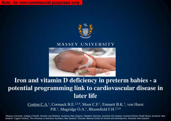

Note: for non-commercial purposes only Iron and vitamin D deficiency in preterm babies - a potential programming link to cardiovascular disease in later life Conlon C.A. 1 , Cormack B.E. 2,3,4 , Moor C.F. 1 , Emmett B.K. 1 , von Hurst P.R. 1 , Mugridge O.A. 1 , Bloomfield F.H. 2,3,4 1 Massey University, College of Health, Nutrition and Dietetics, Auckland, New Zealand, 2 Newborn Services, Auckland City Hospital, Auckland District Health Board, Auckland, New Zealand, 3 Liggins Institute, The University of Auckland, Auckland, New Zealand, 4 Gravida: National Centre for Growth and Development, Auckland, New Zealand
Study location
Study location
Iron and vitamin D deficiency • Reduced accretion, increased requirements for postnatal growth, excessive losses and lack of sun exposure. • Deficiency states are associated with an array of issues for the preterm infant including impaired neurodevelopment, immune dysfunction and bone development. • Also increased risk of iron overload due to immature regulatory mechanisms Can lead to impaired neurodevelopment
Beyond neurodevelopment & bone health • Preterm birth has been associated with increased risk of cardiovascular disease (CVD) in later life. • It has been hypothesised that common micronutrient deficiencies in early life may contribute to CVD risk in preterm babies. • Experimental data suggest that iron and vitamin D deficiency may alter development of the heart, vasculature and metabolic pathways resulting in altered function, thereby potentially leading to cardiovascular disease.
Article Fetal anemia leads to augmented contractile response to hypoxic stress in adulthood. • In sheep, fetal anaemia alters coronary conductance and Craig S Broberg, George D Giraud, Jess M Schultz, Kent L Thornburg, A Roger Hohimer, Lowell E Davis increases susceptibility to Division of Maternal-Fetal Medicine, Medical Research Bldg., L-458, 3181 SW Sam Jackson Park Rd., Portland, OR 97201-3098, USA. ischaemia-reperfusion injury AJP Regulatory Integrative and Comparative Physiology (Impact Factor: 3.28). 10/2003; 285(3):R649-55. DOI:10.1152/ajpregu.00656.2002 in adulthood. Source: PubMed ABSTRACT In response to chronic fetal anemia, coronary blood flow, maximal coronary • This study demonstrates that conductance, and coronary reserve increase. We sought to determine whether chronic fetal anemia alters left ventricular (LV) function in adulthood. We studied adult sheep that had anaemia in utero at an been made anemic for 20 days in utero by phlebotomy. They were transfused just before birth. At 7 mo of age, LV function was measured by pressure-volume loops at rest and during equivalent gestational age as hypoxic stress. The in utero anemia group (n = 8) did not differ from controls (n = 5) with respect to hematocrit, heart and body weight, or baseline hemodynamic parameters. However, preterm babies may be the effect of hypoxia (relative to baseline) on multiple indexes of systolic function was different between the two groups. End-systolic elastance increased in the in utero anemia group (baseline to hypoxia) by 4.15 +/- 3.47 mmHg/ml (mean +/- SD) but changed little in associated with altered controls (0.24 +/- 0.45), which shows that the response to hypoxia was significantly different (P < 0.01) between groups. Similarly, the maximum derivative of LV pressure with respect to cardiac development. time increased in the in utero anemia group (486 +/- 340 mmHg/s,) but on average fell in the controls (-503 +/- 211 mmHg/s) with the response again being significantly different (P < 0.03). We conclude that in sheep, perinatal anemia can alter cardiac responses to hypoxic stress in the adult long after restoration of normocythemia.
• T hese findings provide the first evidence in humans that fetal anaemia is associated with an increased cardiovascular risk profile in adulthood.
Vit D deficient, young male and female offspring have elevated mean arterial pressure (MAP) and heart rate (HR) and endothelial vasodilator dysfunction
Aim • The aim of this study was to determine the iron and vitamin D status of preterm babies after hospital discharge.
Routine supplementation ELBW LBW VLBW <1000g <2500g <1500g On NICU at Auckland only preterm infants <32w and/or <1800g routinely receive 3-6mg/kg/d iron and vitamin D supplements (311-620IU per day) unless clinically indicated
Methods • Situational analysis. • Babies (< 37 weeks ´ gestation) were recruited to the study 4 months after discharge through NICU at Auckland City Hospital. • At four months after discharge measured infant haemoglobin (<110g/L), serum ferritin (<12µg/L) and soluble transferrin receptor (>2.4mg/L), C-reactive protein and serum 25-hydroxyvitamin D (25(OH)D) concentrations (insufficiency ≤50nmol/L). • Anthropometric measures: weight, length and head circumference were measured. • Information about supplementation and mode of feeding was collected.
Contact details retrieved for 208 Study design potential participants from NICU log book 128 Parents could not be 80 Parents Contacted contacted 24 Parents declined 56 Parents consented Infants; n=73 2 infants excluded 4 parents could not be • 1 still in NICU • 1 located outside of Auckland region contacted to make an appointment Eligible Participants n=67 (17 sets of twins) 18 Infants excluded because 25(OH)D biomarkers not available 6 Infants excluded – iron biomarkers not available Final n=61 for iron results n=48 for 25(OH)D
Characteristics of the babies according to receiving or not receiving iron supplements Characteristics P value Infants who received Infants who did not receive supplements after discharge supplements after discharge n= 30 n= 31 Gender n (%) Males 12 (40) 23 (74.2) Females 18 (60) 8 (25.8) 0.007* Gestational Age (weeks+days) Median [25 th , 75 th percentile] 32+5 [30+0, 34+3] 35+3 [34+3, 36+5] <0.001** ≤32 weeks n (%) 14 (46.7) 0 (0) >32 weeks n (%) 16 (53.3) 31 (100) <0.001* Birth Weight (kg) Mean±SD 1.58±0.48 2.54±0.40 <0.001** ≤1.8kg n (%) 23 (76.7) 0 (0) >1.8kg n (%) 7 (23.3) 31 (100) <0.001* Infant Ethnicity n (%) European 17 (56.7) 17 (54.8) Maori 5 (16.7) 3 (9.68) Pacific Island 1 (3.33) 5 (16.1) Asian 3 (10.0) 5 (16.1) Indian 4 (13.3) 1 (3.23) Singleton Birth n (%) 15 (50) 13 (41.9) Twin n (%) 15 (50) 18 (58.1) 0.53
Results Incidence of Iron Deficiency and Iron Deficiency Anaemia in Preterm Infants at Four Months after Discharge Iron Deficiency Iron Optimal Iron Iron Anaemia a Deficiency b Status c Overload d n (%) 10 (16.4) 4 (6.6) 47 (77.0) 0 (0) a Defined as haemoglobin <110 g/L, serum ferritin <12 µg/L and/or sTfR>2.4 mg/L; b Defined as haemoglobin >110 g/L, serum ferritin <12 µg/L and/or sTfR>2.4 mg/L; c Defined as haemoglobin>110 g/L, serum ferritin >12 µg/L and sTfR<2.4 mg/L; d Defined as serum ferritin >300 µg/L
Characteristics of Preterm Infants with Suboptimal Iron Status at Four Months after Discharge Characteristics Optimal Iron Suboptimal Iron P value Status Status Incidence n (%) 47 (77.0) 14 (23.0) - Gestational Age ≤32 weeks n (%) 14 (100) 0 (0) >32 weeks n (%) 33 (70.2) 14 (29.8) 0.026* Birth Weight ≤1.8kg n (%) 20 (87) 3 (13) >1.8kg n (%) 27 (71.2) 11 (28.8) 0.15 Received Iron Supplements Yes n (%) 27 (90) 3 (10) No n (%) 20 (64.5) 11 (35.5) 0.018**
Characteristics of the babies according to receiving or not receiving vitamin D supplements Characteristics P value Infants who received Infants who did not receive supplements after discharge supplements after discharge n= 25 n= 23 Gender n (%) Males 8 (32) 18 (78.3) 0.0001 Females 17 (68) 5 (21.7) 0.0001 Gestational Age ≤ 32 weeks n (%) 11 (44) 0 (0) <0.001** >32 weeks n (%) 14 (56) 23 (100) <0.001* Birth Weight (kg) Mean±SD 1.5±0.44 2.5±0.51 <0.001** ≤1.8kg n (%) 19 (76) 2 (9.5) >1.8kg n (%) 6 (24) 21 (90.5) <0.001* Infant Ethnicity n (%) European 11 (44) 13 (56.5) Maori 2 (8) 1 (4.3) Pacific Island 2 (8) 4 (17.5) Asian 4 (10.0) 1 (4.3) Indian 4 (13.3) 2 (8.7) Singleton Birth n (%) 13 (52) 11 (47.8) Twin n (%) 12 (48) 12 (52.2) 0.53
Feeding Type With or Without Vitamin D Supplements, by Serum 25(OH)D Concentrations in Preterm Infants * * Ŧ * Ŧ n=12 * n=9 n=13 n=14 <0.008 = level of significance, n=number * P=0.0001; Exclusive breastfeeding without Vitamin D supplements versus exclusive breastfeeding with Vitamin D supplements, formula fed without Vitamin D supplements and formula fed with Vitamin D supplements Ŧ P=0.002; Formula fed with Vitamin D versus formula fed without Vitamin D
Conclusion • We conclude that suboptimal micronutrient status in preterm babies 4 months after hospital discharge may be common in babies who don’t receive routine supplementation.
Interpretation • Optimising nutrition after discharge as well as in hospital may ameliorate the potential cardiovascular disease burden in survivors of preterm birth, a population that is increasing steadily worldwide
Thank you • Charlotte Moor, Barbara Cormack, Briar Emmett, Owen Mugridge & Professor Frank Bloomfield. • To all the babies and families who took part in the study
Recommend
More recommend