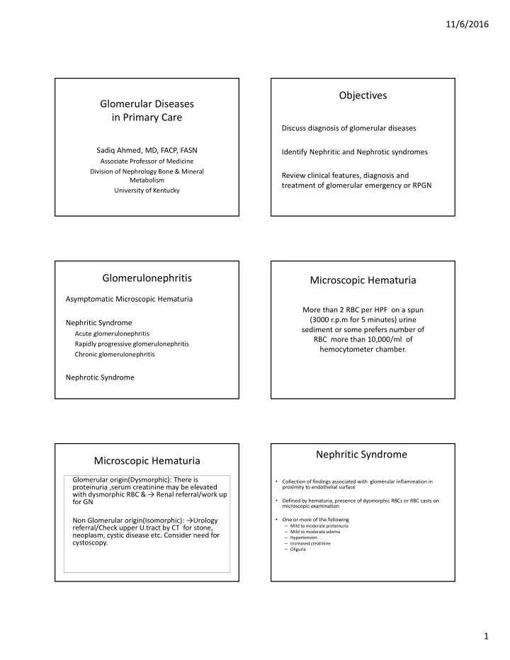

11/6/2016 Objectives Glomerular Diseases in Primary Care Discuss diagnosis of glomerular diseases Sadiq Ahmed, MD, FACP, FASN Identify Nephritic and Nephrotic syndromes Associate Professor of Medicine Division of Nephrology Bone & Mineral Review clinical features, diagnosis and Metabolism treatment of glomerular emergency or RPGN University of Kentucky Glomerulonephritis Microscopic Hematuria Asymptomatic Microscopic Hematuria More than 2 RBC per HPF on a spun (3000 r.p.m for 5 minutes) urine Nephritic Syndrome sediment or some prefers number of Acute glomerulonephritis RBC more than 10,000/ml of Rapidly progressive glomerulonephritis hemocytometer chamber. Chronic glomerulonephritis Nephrotic Syndrome Nephritic Syndrome Microscopic Hematuria Glomerular origin(Dysmorphic): There is Collection of findings associated with glomerular inflammation in • proteinuria ,serum creatinine may be elevated proximity to endothelial surface with dysmorphic RBC & → Renal referral/work up Defined by hematuria, presence of dysmorphic RBCs or RBC casts on for GN • microscopic examination Non Glomerular origin(Isomorphic): → Urology One or more of the following • – Mild to moderate proteinuria referral/Check upper U.tract by CT for stone, – Mild to moderate edema neoplasm, cystic disease etc. Consider need for – Hypertension cystoscopy. – Increased creatinine – Oliguria 1
11/6/2016 Figure 25.7a Clinical differences : Nephrotic vs Nephritic Nephrotic syndrome Nephritic syndrome Proteinuria Gross >3.5GM Moderate < 3GM Serum Albumin Reduced Normal or mild reduction Hematuria Absent or trace Marked Bland urine sediment RBC cast /dysmorphic Active urine sediment Edema Marked Moderate Lipids Marked elevation Minimal elevation /normal Urine volume Normal /reduced Reduced Nephrotic Syndrome Nephritic syndrome Minimal change disease • Post infectious GN FSGS • Lupus Nephritis class II, III,IV Membranous Nephropathy • RPGN Diabetic Nephropathy • Anti ‐ GBM Amyloidosis • Immune complex crescentic GN • Pauci immune. ANCA + crescentic necrotizing GN Class V Lupus Nephritis • IgA nephritis & HSP • MPGN I,II,III 2
11/6/2016 Clinical Clues to Glomerular Diseases Tests for Glomerular Diseases Hematuria, Foamy urine , Elevation of Cr • CBC with diff, Renal panel, UA & Urine Protein and Creatinine Ratio • Pulmonary infection / Infiltrate / Hemoptysis • C3, C4, Ch50 • Hepatitis B, C & HIV infections • ASO titre • Arthralgia • ANA, Anti Double Stranded DNA • Skin Rash • ANCA ( C ‐ ANCA, P ‐ ANCA) • Volume over load / new onset edema • New onset Hypertension • Anti GBM Antibodies • Diseases – Endocarditis, Shunt infection, SLE, • Serum Protein Electrophoresis, Serum Protein IF, Free light chain Lymphoproliferative disorders etc assay • IVDU • Hep ‐ B, C and HIV RPGN Clinical presentation of RPGN Clinical condition that evolve with rapidly progressive decline in renal function and • Rapid loss of renal function characterized by an inflammatory process that • Active urinary sediment results in the formation of cellular crescents & • Oliguria called crescentic glomerulonephritis. • Hematuria/Proteinuria This is glomerular emergency. RPGN How Crescents are formed? Glomerular crescent formation is an etiologically nonspecific response to glomerular capillary rupture due to acute inflammatory injury • Rupture of GBM → spillage of in fl am. mediators • Neutrophil margination • Karyorrhexis • Thrombosis • Fibrin exudation • Epithelial response 3
11/6/2016 RPGN:Case Electron microscopy of RPGN 65 YO W male presents to the ER with 3 weeks history of arthralgia and fatigue & URI. On exam. BP is 150/80 and there is a petechial rash visible in lower extremities and also 1+ edema. Hb is 9.4gm/dl, Creatinine 5.8mg/dl Urinalysis shows 20 ‐ 50 RBC/hpf & 1+ protein CxR showed bilateral infiltrate. 3 major Immunopathologic categories RPGN:Case of crescentic GN What is the most likely cause of his renal failure? • Type I A. Anti GBM disease – Anti ‐ GBM crescentic GN B. Lupus nephritiis • Type II C. Henoch Schoenlein – Immune complex crescentic GN purpura with crescentic • Post infectious, SLE, IgA, MPGN, Fibrillary GN • Type III D. Cryoglobulinemia – Pauci ‐ immune crescentic GN E. ANCA associated – 80% ANCA positive vasculitis Crescentic GN and Systemic Vasculitis ANCA test methodology • Iimmunofluroscnce technique 75% of patients with ANCA GN have some sort of – On ethanol fixed neutrophils PR3 ‐ ANCA causes a systemic small vessel vasculitis. GPA,MPA or EGPA charecteristic cytoplasmic granular centrally accentuated immunofluroscence pattern called cANCA while MPO ‐ ANCA causes a perinuclear pattern called pANCA 50 ‐ 60% patients with Anti GBM disease have • Antigen specific testing pulmonary cappillaritis – Antigen type (PR3 or MPO) is determined through antigen ‐ specific methods. More specific • ELISA or capture ELISA Immune complex crescentic GN has the lowest • Bead based multiplex assay frequency of systemic vasculitis. HSP, • Appropriate paring is important for definitive DX Cryoglobulinemic GN etc. – cANCA with PR3 ‐ ANCA & pANCA with MPO ‐ ANCA 4
11/6/2016 Differential Dx of ANCA disease C ‐ ANCA. Cytoplasmic granular centrally P ‐ ANCA. Perinuclear Imm. fluoroscent Cleveland clinic journal of Medicine Supp.3,V 79, Nov.2012 accentuated Imm.fluoroscent pattern ‐ pattern ‐ MPO PR3 GPA MPA EGPA ENT Necrotizing None Allergic Lung Nodule, Cavity, Infiltrate Allergy, Asthma Infiltrate Kidney ++++ ++++ ++ Nerve ++ +++ ++++ Granuloma +++++ None ++++ Eosinophil None None ++++ Skin ++ ++++ ++++ ANCA 80 ‐ 95% PR3 40 ‐ 80% MPO 40% MPO 35% PR3 35% PR3 5 ‐ 20% MPO 0 ‐ 20% negative 0 ‐ 20% negative Up to 60% negative Types of Crescentic GN in consecutive Renal Treatment of ANCA Vasculitis Biopsies in Univ. of North Carolina • Pulse steroids 1 GM daily for 3 days followed by Prednisone 1 mg / Kg daily • Cyclophosphamide or Rituximab • Plasmapheresis • Diffuse Alveolar hemorrhage • Severe Renal failure RPGN Type I: Goodpasture’s RPGN Type I: Goodpasture’s Syndrome Syndrome Anti ‐ GBM antibody ‐ induced disease. • The anti ‐ GBM antibodies cross ‐ react with pulmonary alveolar Cells accumulate in Bowman’s space, form • basement membranes to crescents . produce the clinical picture of pulmonary hemorrhage The Goodpasture antigen is a peptide within the • noncollagenous portion of the α 3 ‐ chain of associated with renal failure. collagen type IV. What triggers the formation of these antibodies • is unclear There is linear deposition of antibodies and • complement components along the GBM. 5
11/6/2016 Frequency of crescents in different types of glomerular Treatment of Good Pastures Syndrome diseases in University of North Carolina. Kidney International, Vol 63 (2003) pp.1164 ‐ 1177 Numbers % with any % with >50% Average % of • Plasmapheresis daily ASAP crescents crescents crescents Anti ‐ GBM 105 97.1 84.8 77 ANCA GN 181 89.5 50.3 49 • Pulse Steroids 1 GM daily for 3 days and then 1 Lupus ‐ III & IV 784 56.5 12.9 31 mg / Kg daily HSP 31 61.3 9.7 27 IgA 853 32.5 4 21 Post ‐ infectious 120 33.3 3.3 19 • Cyclophosphamide IV or PO Type I MPGN 307 23.8 4.6 25 Type II MPGN 16 43.8 18.8 48 Fibrillary GN 101 22.8 5 26 Mon. Ig dep. dx 54 5.6 0 13 TMA 251 5.6 0.9 20 Membranous 1092 3.2 0.1 15 RPGN IgA nephropathy Which type of glomerulonephritis usually do not present as RPGN ? Autoimmune systemic disease –kidneys are bystanders A. IgA nephritis B. Lupus nephritis Defective glycosylation of IgA1 fraction with with C. Fibrillary GN galactose deficiency D. FSGS 75% of patients with IgA nephropathy has galactose deficient IgA1 level above 90 th percentile Glomerular and tubular injury by immune complexes containing containing pathogenic IgA1 Watt Rj, Julian BA, New Eng. J Med 2013;368;2402 ‐ 2414 6
Recommend
More recommend