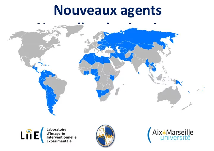

Nouveaux agents Nouvelles destinations Fair-Embolisation et agents liquides Vincent VIDAL
Nouveaux liquides Onyx, Squid, Phil, Easyx and others Transcatheter embolization in animal models with GPX, a novel water-borne polymer embolic. Joshua P. Jones, PhD; Jessica C. Karz; and Matthew S. Johnson, MD, FSIR Diverse Use Scenarios Purpose Results • When injected proximal to coils in branches of the iliac, splenic, and GPX (Fluidx Medical Technology, Salt Lake City, UT) is a Swine Kidney and Liver Embolization renal arteries, GPX formed a new type of transcatheter embolic: a water-based • GPX distally penetrated into small arteries cohesive embolus that did not coacervate which solidifies upon injection into the without fragmentation, resulting in Initial Minimal With GPX: total Coil placement travel distal to the coils (Fig. 3). vasculature. The agent is primarily comprised of two Initial angiogram GPX placement angiogram occlusion occlusion immediate occlusion. • In an acute procedure, GPX oppositely charged polymers, with interactions • All treatment sites [n=20] showed no flow Figure 3: Diverse uses of demonstrated deep penetration shielded by high concentrations of monovalent ions (AFA grade 0) at the acute follow-up, GPX. Top: With coil. Right: into the rete mirabile, resulting in (salt). Tantalum powder is added for radiopacity. Acute necropsy. Bottom: compared with an average of 0.7 (+/-0.6) rapid complete occlusion with no Swine rete mirabile (left) Solidification results from strengthened interactions for microspheres [n=4]. signs of crossing into venous and aneurysm model between the oppositely charged polymers in • In the survival arm, all sites remained fully Necropsy minutes post-injection: (right). circulation (Fig. 3). GPX did not fragment and response to decreasing salt concentration (Figure 1). 15 minutes post 14 days post occluded (AFA=0) at 14 days [n=7]. displayed distal penetration • GPX totally occluded model wide- This produces a cohesive viscoelastic gel that can be Microspheres had an average AFA score of Figure 2: neck aneurysms at both acute delivered with control and precision without adhering 2.3 (+/- 1.2) [n=4]. Porcine kidney and 1 month evaluations with no to catheters. Here, the performance of the GPX • At necropsy, GPX remained cohesive and embolization signs of catheter or balloon embolic was evaluated in procedures out to 14 days was well distributed in the target tissue. using GPX at adhesion. Explanted tissue in a porcine model and 1 month in a rabbit model. 14 days. Penetration was seen into small arterioles. Initial GPX No balloon adhesion after appeared grossly normal (Fig. 3). Faxitron of GPX angiogram GPX was used alone and in combination with other embolization embolization of rabbit aneurysm agents to simulate a variety of clinical scenarios. AFA Flow Scale Materials & Methods Conclusions Grade Definition AFA Swine Kidney and Liver Embolizations • When used as a stand-alone embolic, GPX Grade No antegrade flow within the treatment site. All procedures/evaluations were done by non-Fluidx personnel. • 0 demonstrated good distal penetration, filling down to A Boston Scientific Direxion HI-FLO microcatheter (0.027” ID) • AFA Contrast passes into the treatment site but “hangs the arterioles, and generating a stable occlusion. was placed proximal to the target vasculature. up” and fails to opacify the entire distal vascular Grade GPX was injected (~0.3 mL) over 1 min. Embozene Microspheres • • In the porcine kidney model, kidney lobes embolized bed. 1 (40µm) were used as a control (deployed as per the IFU). with GPX had less flow at the acute and long term Antegrade filling of contrast with complete filling of Occlusion was assessed at 15 m and 14 d (see Table 1). • the artery and its major and minor branches after follow-up than those performed with Embozene AFA more than two cardiac cycles. Alternatively, Diverse Use Scenarios Microspheres (40 µm). Grade delayed contrast washout in the target site territory GPX was deployed into coils at multiple sites including the rete • 2 may occur , compared with comparable areas of • When used in tandem with coils, the cohesiveness of mirabile as well as the splenic, gastric, and renal arteries. distal vascular bed not perfused by the target site. GPX allows it to form a proximal, complete occlusion In rabbits, elastase was used to create model aneurysms in the • Antegrade flow of contrast with complete filling of without distal migration in different vessel/flow subclavian artery. Embolization was performed using a balloon AFA the vessel and its major and minor branches within Coacervate Post Injection Grade and jailed catheter technique. Follow-up was performed at 28 d. situations. two cardiac cycles. Contrast also clears from the 3 (6-9% Saline) (0.9% Saline) These procedures explored the use of GPX with a variety of • arterial segment within two cardiac cycles. • GPX is a flow directed embolic agent that appears to catheters ranging from 0.017” ID (Medtronic Echelon 10) to Table 1: The AFA Flow Scale was used to grade Figure 1: GPX solidifies after delivery in have utility in a variety of embolization scenarios. 0.041” ID (Terumo Glidecath). occlusion at 15 minutes and 14 days. response to decreasing salt concentration.
GPX • Polymeres à charge opposées + Tantalum • Retenus par une barrière d’ions monovalents • Diminution de la concentration Coacervate Post Injection (6-9% Saline) (0.9% Saline) Figure 1: GPX solidifies after delivery in response to decreasing salt concentration.
Initial angiogram GPX placement 15 minutes post 14 days post
With GPX: total Initial Minimal Coil placement occlusion angiogram occlusion Figure 3: Diverse uses of GPX. Top: With coil. Right: Acute necropsy. Bottom: Swine rete mirabile (left) and aneurysm model Necropsy minutes post-injection: (right). GPX did not fragment and displayed distal penetration Initial GPX No balloon adhesion after angiogram embolization embolization of rabbit aneurysm
Nouveaux liquides Onyx, Squid, Phil, Easyx and others • GPX • Polymeres + solvant • Radio-opacité • Viscosité • Rupture technogique …
The Fair-Embo Concept Faisabilité in vivo de l’embolisation artérielle avec des fils de sutures
Radiologie Interventionnell e
PRIX
DISPONIBILITE DISTRIBUTION
The Fair-Embo Concept • Promouvoir : • RI dans les pays émergents • Développement d’un agent d’embolisation : • Efficace • Disponible • Peu couteux
Matériels & méthodes : Ex VIVO 13
Matériels & méthodes : Ex VIVO 14
Materiels & méthodes : In VIVO • AIGU : (n = 1) • Sécurité (hors cible) • Succès technique • mesenterique, splenique, pharyngée, rénale • Suture non-absorbable (SNA): • Mersutures T M 6, Ethicon (polyethylene terephthalate) • diamètre 0.8 and 0.899 mm, préparé en fragments de 1 à 3 cm
Materiels & méthodes : In VIVO • C hronique (n = 3) : • efficacité et tolerance ( moyen terme) • Artère polaire rénale inférieure : • Droite : SNA • Gauche : SA Vicryl, polyglactin 910: Ethicon. • Suivi 3 mois . • Suture taille 1 • diametre USP • 0.4 / 0.5 mm, fragments de 1 cm (3 mg)
Materiels & méthodes : In VIVO Protocole J-1 J0 M1 M3 CT- Scanner X X X X Angiographie X X X Histological study
Résultats : In VIVO • AIGU : • Securité • Succès technique
Résultats : In VIVO • AIGU : • Polyethylene terephthalate • 6 USP (0.8–0.9 mm) 4 Fr (0.97 mm) • Variation possible 1 à 3 cm voir plus • Occlusion rapide avec 2 à 4 fragments (9.3 ± 3.4 cm).
Résultats : In VIVO • CHRONIQUE : • Rein Dt : SNA , polyethylene terephthalate • 1 cm USP 1 : 21.3 ± 5.6 fragments (0.064 g ± 0.017, n = 3). • Rein Gch : SA , polyglactin 910 • 1 cm USP 1 : 17.7 ± 4.6 fragments (0.053 g ± 0.014, n = 3).
Résultats : In VIVO • Angiographie à 3 mois SNA SA J0 M3 J0 M3 n ° 1 n ° 2 n ° 3
Résultats : In VIVO • Scanner à 1 et 3 mois : • Pas de complications (abces, urinome). • 3 mois, • Atrophie polaire inférieure
Résultats : In VIVO • SNA • Matériel non fragmenté retrouvé dans tous les reins droits, rien à gauche (SA) !
Résultats : In VIVO • SA totalement résorbé à 3 mois • Pas d’infiltration perivasculaire. • no significant difference of tissue fibroplasia, edema, neovascularization, hemorrhage, necrosis, or polymorph nuclear cells, lymphocytes, plasma cells, macrophages and giant cells observed in all the sections.
PUBLICATION CVIR April 2019
DISCUSSION
DISCUSSION
DISCUSSION • Avantages • Low cost • Disponibilité universelle • Facile d’utilisation (taille, flushable) • Toxicité (implantation) • Désavantages • Linéaire (accrochage, torpille gélatine) • Non radio-opaque (particules et gelatine)
Recommend
More recommend