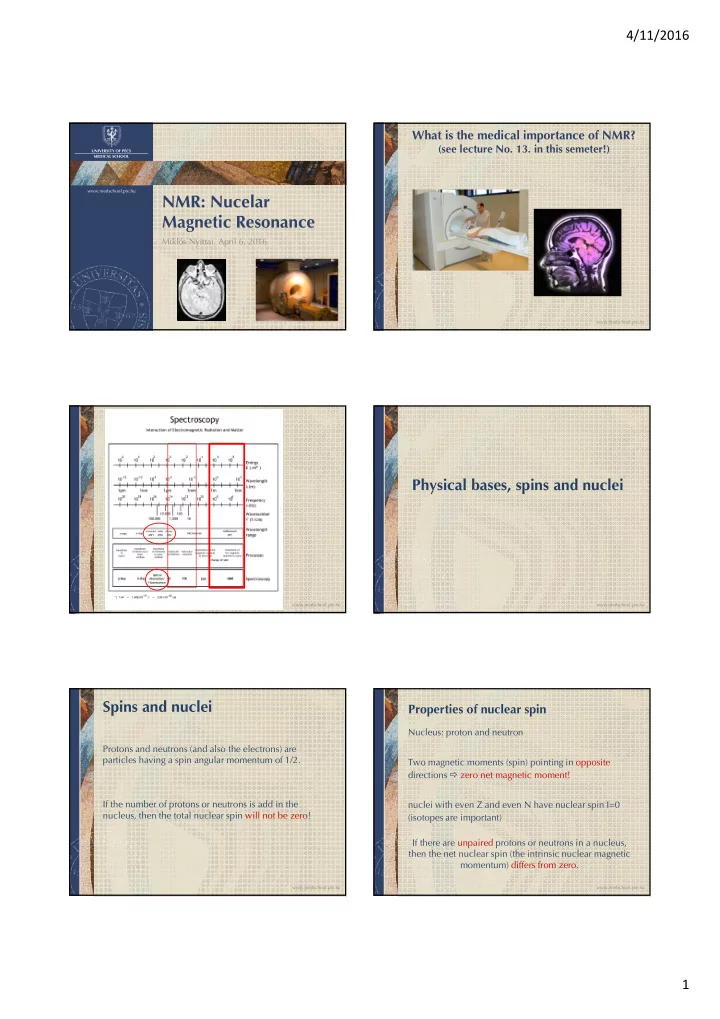

4/11/2016 What is the medical importance of NMR? (see lecture No. 13. in this semeter!) UNIVERSITY OF PÉCS MEDICAL SCHOOL www.medschool.pte.hu NMR: Nucelar Magnetic Resonance Miklós Nyitrai, April 6, 2016 www.medschool.pte.hu Physical bases, spins and nuclei www.medschool.pte.hu www.medschool.pte.hu Spins and nuclei Properties of nuclear spin Nucleus: proton and neutron Protons and neutrons (and also the electrons) are particles having a spin angular momentum of 1/2. Two magnetic moments (spin) pointing in opposite directions � zero net magnetic moment! If the number of protons or neutrons is add in the nuclei with even Z and even N have nuclear spin I=0 nucleus, then the total nuclear spin will not be zero! (isotopes are important) If there are unpaired protons or neutrons in a nucleus, then the net nuclear spin (the intrinsic nuclear magnetic momentum) differs from zero. www.medschool.pte.hu www.medschool.pte.hu 1
4/11/2016 The split of the energy states (Zeemann) Atoms for NMR 1 H, 2 H, 3 H, 3 He, 4 He, 12 C, 13 C, 14 C, 14 N, 15 N 16 O, 17 O, 19 F, 23 Na, 31 P, etc. N N β -spin state, α -spin state, The most frequently applied nuclei: 1 H, 13 C, 15 N, 17 O, 19 F, F, 31 P unfavourable, favourable, lower energy higher energy N S S N S S Signal depends on: Spinning nucleus with it’s magnetic field Spinning nucleus with it’s magnetic field alligned with the external field. alligned against the external field. - Magnitude of the magnetic moment The energy difference between the spin states does depend on the - Concentration of the isotope strength of the external magnetic field. www.medschool.pte.hu www.medschool.pte.hu Orientation and reorientation on the Graphical representation microscopic level M= Σ µ i N E N at no magnetic field there β -spin is no difference between these states state S S µ µ µ ∆ E 1 < ∆ E 2 µ µ N S α -spin state N < H 1 H 2 H S H=0; M=0 H>0; M>0 Remember, the energy is proportional to the frequency! m: magnetic moment of the individual atom www.medschool.pte.hu www.medschool.pte.hu An example: CH 4 What is the problem with this concept? E β -spin state h x 300MHz h x 500MHz It is difficult to compare the data from two instruments with different strenghts of magnetic fields. α -spin state Let’s use the chemical shift! What is this? Much simpler than it may sound… H 0 T 11,75 T 7,05 T We can probe the energy difference of the α - and β - state of the protons by irradiating them with EM radiation of just the right energy. In a magnet of 7.05 Tesla, it takes EM radiation of about 300 MHz (radio waves). So, if we bombard the molecule with 300 MHz radio waves, the protons will absorb that energy and we can measure that absorbance. In a magnet of 11.75 Tesla, it takes EM radiation of about 500 MHz (stronger magnet means greater energy difference between the α - and β - state of the protons). www.medschool.pte.hu www.medschool.pte.hu 2
4/11/2016 An example: chemical shift Chemical shift or δ Imagine that we have a magnet where our standard absorbs at 300,000,000 Hz (300 megahertz), and our sample absorbs at We need a reference sample to standardize our insturments! 300,000,300 Hz. The difference is 300 Hz, so we take 1. Measure the absorbance frequency of the standard: f r ; 300/300,000,000 = 1/1,000,000 and call that 1 part per million (or 1 2. Measure the frequency for your own sample: f s ; PPM). 3. Let’s calculate the difference between the two: ∆ f = f r - f s ; 4. Normalise the difference with the ferquency of the reference: δ = ∆ f / f r ; Now lets examine the same sample in a stronger magnetic field where the reference comes at 450,000,000 Hz, or 450 megahertz. The frequency of our sample will increase proportionally, and will come at 450,000,450 Hz. The difference is now 450 Hz, but we divide by The δ, i.e. the chemical shift will be characteristic for your 450,000,000 (450/450,000,000 = 1/1,000,000, = 1 PPM). sample, but will not depend on the parameters of the instrument used for the experiments! (We do not have to calculate all these, the NMR machine does it for us!) Why? Let’s take an example! www.medschool.pte.hu www.medschool.pte.hu NMR spectrum Applications of NMR Structure of organic molecules and subtances; The absorption is proportional to the concentration of the corresponding nuclei. Interactions of organic molecules; The NMR spectrum is the energy absorbed by the Structure of macromolecules (proteins, nucleic acids); system as the function of the freqeuncy (f) of the excitation energy ( Δ E) or the magnetic field (H, B). Biological and artificial membranes; Due to local effects and fields the excitation energy (and MRI: Magnetic Resonance Imaging. thus frequency) is different for different nuclei. www.medschool.pte.hu www.medschool.pte.hu www.medschool.pte.hu www.medschool.pte.hu 3
4/11/2016 Jablonski diagram SUMMARY Excited-state D A Vibrational relaxation – heat (10 -12 s) S 1 → S 1 → T 1 : inter- k t system crossing (10 -10 – 10 -8 s) T 1 excitation (10 -15 s) S 1 → S 0 (10 -9 s ) S 0 – S 1 T 1 → S 0 (10 -3 – 10 -1 s ) h ν 5 4 3 2 S 0 1 0 vibrational Ground-state levels www.medschool.pte.hu www.medschool.pte.hu The definition of absoprtion How to measure absorption? A scheme of a photometer. I 0 I light source monochromator sample detector substance OD = A = - log (I / I 0 ) = ε ( λ ) c x I = I 0 10 - ε ( λ ) c x „optical density” www.medschool.pte.hu www.medschool.pte.hu The absorption of proteins The absorption of proteins www.medschool.pte.hu www.medschool.pte.hu 4
4/11/2016 Definition of fluorescence and phosphorescence Kasha’s-rule S S In the ns range Michael Kasha December 6, 1920 - June 12, 2013 In the > ms range T S The emission of the fluorescence light is allways starting from the lowes est vibrational level of the first excited level (S 1 ). www.medschool.pte.hu www.medschool.pte.hu Stokes-shift Basic fluorescence parameters The differ eren ence ce (measured in nm ) between the peak of the excitation and the emission spectrum (energy loss). � Fluorescence spectrum, intensity; � Quantum efficiency Stokes � Fluorescence lifetime fluorescence excitation sp. fluorescence emission sp. � Polarisation wavelength (nm) www.medschool.pte.hu www.medschool.pte.hu Scheme of a spectrofluorometer Photoselection light source excitation monochromator F M S sample holder r 90 o E emission M monochromator D detection Non linear arrangement !!! www.medschool.pte.hu www.medschool.pte.hu 5
4/11/2016 Scheme of a spectrofluorometer Emission anisotropy light source excitation r = (I VV - GI VH ) / (I VV + 2GI VH ) monochromator F M S sample P Vertica cally y align gned holder Horizo zontal ally align gned ed polar arizer er on the polar arizer er on the G = I HV / I HH excitat ation side emission side P 90 o • dimensionless emission M monochromator • depends on rotational motion of the fluorophore Vertical or horizontal polarisers: • additive! I VV I VH Remember! I sum = I Z + I X + I Y D I HV I HH detection I sum = I VV + I VH + I VH I sum = I VV + 2I VH Polarisers in the light paths!!! www.medschool.pte.hu www.medschool.pte.hu Conditions of FRET FRET efficiency E = 1 – (F DA / F D ) • A fluores escen cent donor molecule. • The appropriate orien entat ation of the donor and where • Over verlap ap between the donor emission and acceptor absorption spectra. F DA : donor intensity with the acceptor • Distance range of 2-10 nm! F D : a donor intensity without the acceptor. Can also be calculated with lifetimes! E = 1 – ( τ DA / τ D ) www.medschool.pte.hu www.medschool.pte.hu Applications of FRET Distance dependence of the FRET efficiency • The determination of FRET distances → To study the establishment of interactions between molecules; → To study intra-molecular structural changes. 6 R 0 = E 6 6 R + R 0 Determination of distances! Molecular ruler! www.medschool.pte.hu www.medschool.pte.hu 6
4/11/2016 Vibration modes of water Model for molecular vibrations molecules spring = bond mass = atom f ~ D / m http://www1.lsbu.ac.uk/water/vibrat.html if bond strength ↑ then f ↑ if m atom ↑ then f ↓ (C=H: 1650 cm -1 , C≡C: 2200 cm -1 ) (C-H: 3000 cm -1 , C-Cl: 700 cm -1 ) www.medschool.pte.hu www.medschool.pte.hu Example: regions of an IR spectrum Infra and Raman spectroscopy Vibrational levels S 1 hf 0 h(f 0 -f 1 ) h(f 0 +f 1 ) Vibrational levels S 0 Rayleigh Bonds to H Triple Double ”Fingerprint” IR abs. Raman scatter scatter (X–H) bonds bonds region (4000-2500 cm -1 ) (2500-2000) (2000-1500) (1500 - 600) elastic vs. non-elastic www.medschool.pte.hu www.medschool.pte.hu Orientation and reorientation on the Graphical representation microscopic level M= Σ µ i N E N at no magnetic field there β -spin is no difference between these states state S S µ µ µ ∆ E 1 ∆ E 2 < µ µ N S α -spin state N < H 1 H 2 H S H=0; M=0 H>0; M>0 Remember, the energy is proportional to the frequency! m: magnetic moment of the individual atom www.medschool.pte.hu www.medschool.pte.hu 7
Recommend
More recommend