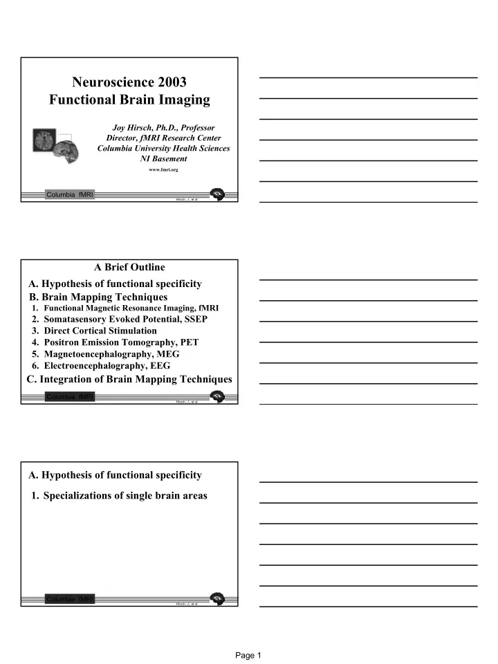

Neuroscience 2003 Functional Brain Imaging Joy Hirsch, Ph.D., Professor Director, fMRI Research Center Columbia University Health Sciences NI Basement www.fmri.org Columbia fMRI Hirsch, J., et al A Brief Outline A. Hypothesis of functional specificity B. Brain Mapping Techniques 1. Functional Magnetic Resonance Imaging, fMRI 2. Somatasensory Evoked Potential, SSEP 3. Direct Cortical Stimulation 4. Positron Emission Tomography, PET 5. Magnetoencephalography, MEG 6. Electroencephalography, EEG C. Integration of Brain Mapping Techniques Columbia fMRI Hirsch, J., et al A. Hypothesis of functional specificity 1. Specializations of single brain areas Columbia fMRI Hirsch, J., et al Page 1
7 7 6 6 Primary Flashing Visual Cortex LED Display 5 5 4 4 Calcarine Sulcus Columbia fMRI Hirsch, J., et al Harrington, 1964 R Previous lesion Surgical lesion Visual Field BINOCULAR FLASHING LIGHTS Left Eye Right Eye Columbia fMRI Hirsch, J., et al HISTORICAL MILESTONES 1841 1874 1890 1909 1950 BROCA WERNICKE ROY & BRODMANN PENFIELD SHERRINGTON Intraoperative Aphasia and Aphasia and Relationship Cytoarchitectonic cortical maps lesions in lesions in between neural regions of cortex GFi GTs activity and vascular changes 1845 1936 1945 1971 PAULING PURCELL HOUNSFIELD FARADAY Magnetic state of BLOCK Magnetic properties CORMACK hemoglobin changes of blood Demonstrated with oxygenation Invention of NMR in condensed Computed RABI matter Tomography Discovery of Magnetic Resonance Columbia fMRI Hirsch, J., et al Page 2
Damasio, H., et al; Science 264: 1102-1105, 20 May 1994 Columbia fMRI Hirsch, J., et al HISTORICAL MILESTONES 1990 1977 OGAWA TER-POGOSSIAN SOKOLOFF 1992 fMRI First PET studies of brain BELLIVEAU metabolism, blood flow, and correlates of human behavior Cortical Map: Blood Oxygen human visual 1971 1972 1976 dependent signal system EPI / MRI Behavior MANSFIELD DAMADIAN LAUTERBUR First MRI of a body part Discovered that First MR image biological tissues Invented EPI (scans whole have different brain in secs.) relaxation rates Columbia fMRI Hirsch, J., et al A. Hypothesis of functional specificity 1. Specializations of single brain areas 2. Specializations of multiple brain areas Columbia fMRI Hirsch, J., et al Page 3
Functional Organization of Visual Cortex Columbia fMRI Hirsch, J., et al B. Brain Mapping Techniques 1. Functional Magnetic Resonance Imaging, fMRI Columbia fMRI Hirsch, J., et al Early Developments in MR Solved the Occluded-Structure Problem Sadek K. Hilal, M.D., Ph.D. Department of Radiology Columbia University Columbia fMRI Hirsch, J., et al Page 4
L R Columbia fMRI Hirsch, J., et al Current Developments in MR are focused on the structure/function problem Left Hand - Touch MRI Signal Intensity L R REST TASK REST REST TASK REST - 40 s - - 40 s - - 40 s - - 40 s - - 40 s - - 40 s - 2 minutes 24 seconds From Hirsch, J., et al; An Integrated Functional Magnetic 2 minutes 24 seconds Resonance Imaging Procedure for Preoperative Mapping of TIME Cortical Areas Associated with Tactile, Motor, Language, and Visual Functions, Neurosurgery 47: 711-722, 2000. Columbia fMRI Hirsch, J., et al B. Brain Mapping Techniques 1. Functional Magnetic Resonance Imaging, fMRI a. Source of signal Columbia fMRI Hirsch, J., et al Page 5
PHYSIOLOGY PHYSICS NEURAL ACTIVATION DEOXY HGB IS ASSOCIATED WITH AN IS PARAMAGNETIC INCREASE IN BLOOD FLOW O2 EXTRACTION IS AND DISTORTS THE LOCAL RELATIVELY UNCHANGED MAGNETIC FIELD CAUSING SIGNAL LOSS RESULT: RESULT: REDUCTION IN THE LESS DISTORTION OF THE PROPORTION OF DEOXY HGB MAGNETIC FIELD RESULTS IN IN THE LOCAL VASCULATURE LOCAL SIGNAL INCREASE Columbia fMRI Hirsch, J., et al B. Brain Mapping Techniques 1. Functional Magnetic Resonance Imaging, fMRI a. Source of signal b. Measuremnet techniques Columbia fMRI Hirsch, J., et al Imaging While Naming Objects Columbia fMRI Hirsch, J., et al Page 6
Block Design Event-Related Design Columbia fMRI Hirsch, J., et al B. Brain Mapping Techniques 1. Functional Magnetic Resonance Imaging, fMRI a. Source of signal b. Measuremnet techniques c. Computation for analysis Columbia fMRI Hirsch, J., et al COMPUTATIONS FOR f UNCTIONAL IMAGE PROCESSING RECONSTRUCTION ALIGNMENT VOXEL BY VOXEL Functional Scanner ANALYSIS Brain Map GRAPHICAL Language REPRESENTATION Primary Auditory Cortex Columbia fMRI Hirsch, J., et al Page 7
Run 1 Run 2 MULTI-STAGE ANALYSIS (90 secs) (90 secs) WITH COINCIDENCE Stage 1 COINCIDENCE Run 1 AND Run 2 Stage 2 Penfield’s Motor Homonculus 1 and 2 Left Hand: Finger Thumb Tapping Columbia fMRI Hirsch, J., et al from Nature 388 , 171-174 (1997) Broca’s Area Kim, Relkin, Lee, & Hirsch “LATE” BILINGUAL “EARLY” BILINGUAL (Separate Language Areas) (Overlapping Language Areas) + + + + + + + + R R 0 0 Native (English) Native 1 (Turkish) Second (French) Native 2 (English) + Center-of-Mass Common Region + Center-of-Mass Columbia fMRI Hirsch, J., et al B. Brain Mapping Techniques 1. Functional Magnetic Resonance Imaging, fMRI a. Source of signal b. Measuremnet techniques c. Computation for analysis d. Individual brain maps Columbia fMRI Hirsch, J., et al Page 8
Conventional Functional Imaging Imaging Before Surgery Tumor Tumor R Left Hand: Sensory/Motor CC 23 (AB) Columbia fMRI Hirsch, J., et al Conventional Imaging Tumor R CC 23 (AB) Columbia fMRI Hirsch, J., et al Conventional Functional Imaging Imaging Before Surgery After Surgery Tumor Tumor R Left Hand Left Hand: Sensory/Motor Movement CC 23 (AB) Columbia fMRI Hirsch, J., et al Page 9
Standard Brain Mapping Tasks SENSORY MOTOR LANGUAGE Touch Finger Thumb Picture Listening Naming to Words Tapping (passive) (active) (active) (passive) GPoC GPrC GOi GTT GFi GTs From Hirsch, J., et al; Neurosurgery 47: 711-722, 2000 Columbia fMRI Hirsch, J., et al Functional Organization of Visual Cortex QuickTime™ and a Video decompressor are needed to see this picture. Columbia fMRI Hirsch, J., et al B. Brain Mapping Techniques 1. Functional Magnetic Resonance Imaging, fMRI 2. Somatasensory Evoked Potential, SSEP 3. Direct Cortical Stimulation Columbia fMRI Hirsch, J., et al Page 10
Conventional functional mapping in the OR SSEP - S omatoSensory E voked P otentials Cortical Stimulation Based on Electrical Activity of Neurons Induced by External Stimulation Columbia fMRI Hirsch, J., et al Sensory Motor Mapping Direct Cortical Craniotomy Localization fMRI SSEP Stimulation “Twitching of hand, focal seizure involving arm ” Tag 3 Tag 3 Tag 5 “Twitching in 1st three digits” From Hirsch, J., et al; An Integrated Functional Magnetic Resonance Imaging Tag 5 Procedure for Preoperative Mapping of Cortical Areas Associated with Tactile, Motor, Language, and Visual Functions, Neurosurgery 47: 711-722, 2000. Columbia fMRI Hirsch, J., et al Homonculus Columbia fMRI Hirsch, J., et al Page 11
Language Mapping Intraoperative Response fMRI Stimulation Broca’s Area Speech Arrest During Counting Wernicke’s Area Literal paraphasic speech error during picture naming From Hirsch, J., et al; An Integrated Functional Magnetic Resonance Imaging Procedure for Preoperative Mapping of Cortical Areas Associated with Tactile, Motor, Language, and Visual Functions, Neurosurgery 47: 711-722, 2000. Columbia fMRI Hirsch, J., et al B. Brain Mapping Techniques 1. Functional Magnetic Resonance Imaging, fMRI 2. Somatasensory Evoked Potential, SSEP 3. Direct Cortical Stimulation 4. Positron Emission Tomography, PET a. Source of signal Columbia fMRI Hirsch, J., et al Positron Emission Tomography Radionuclides that emit positrons such as 15 O and 18 F are introduced into the brain. H 2 15 O behaves like H 2 16 O and indicates blood flow (rCBF) (half life = 123 seconds) integration time ≈ 60 seconds. 18 F – deoxyglucose behaves like deoxyglucose and indicates metabolic activity (half-life = 110 PET SCANNER minutes) integration time ≈ 20 minutes From: www.epub.org.br/cm/n011pet/pet.htm Columbia fMRI Hirsch, J., et al Page 12
Principle of PET A 2 Positron and electron annihilation and emission of gamma rays PET is based on the radioactive decay of positrons from the nucleus Gamma ray of the unstable atoms ( 15 O has 8 protons and 7 neutrons) Site of positron annihilation A 1 Positron emission Electron (imaged point) in the brain Gamma ray 0-9mm Unstable Positron photon resolution radionuclide limit From: Principles of Neural Science (4th. Ed.) Kandel, Schwartz, & Jessell, p. 377. Columbia fMRI Hirsch, J., et al B. Brain Mapping Techniques 1. Functional Magnetic Resonance Imaging, fMRI 2. Somatasensory Evoked Potential, SSEP 3. Direct Cortical Stimulation 4. Positron Emission Tomography, PET a. Source of signal b. Measurement techniques Columbia fMRI Hirsch, J., et al Gamma Ray Detections to Location of Function From: Principles of Neural Science (4th. Ed.) Kandel,Schwartz, & Jessell, p. 377. Columbia fMRI Hirsch, J., et al Page 13
Recommend
More recommend