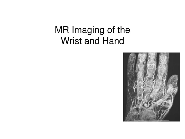

MR Imaging of the Wrist and Hand
MR wrist and hand • Technical considerations • Internal derangement of the wrist – TFCC – Ligaments • Osseous abnormalities • Arthritis, Tendons, and Ligaments • Miscellaneous
Technique • Supine, hand by side (avoid excessive pronation) • Prone, hand above head • Decubitus, hand in front directed cranially • Comfortable immobilization
Protocol • Routine protocol • Tailored protocol for specific indications (tumor, infection) • MR arthrography
Protocol Plane Sequence TR/TE FOV Matrix Slice/ NEX Gap Localizer FMPIR 2800/30 14 128 4/1 1 TI 140 Coronal PD FSE 2500/19 8 256 3/1 2 Coronal T2 FSE 2500/80 8 256 3/1 2 Coronal T2* GE 450/15 8 192 .6 mm 2 30 degree flip Axial PD FSE 2500/19 8 256 3/1 2 Axial T2 FSE 2500/80 8 256 3/1 2 Sagittal T1 SE 600/20 8 256 4/1 1
Imaging planes • Axial sequence done first • Radial styloid to ulnar styloid • Parallel to volar surface of radius
Wrist Arthrography Indications • Intercarpal ligaments • Triangular fibrocartilage • Scaphoid nonunion • Soft tissue ganglia • Wrist prosthesis TFCC and LT ligament perforations
Wrist Arthrography Technique • Controversy about which compartments and how many compartments need to be injected • Most common single injection is radiocarpal Lunotriquetral perforation
Wrist Arthrography Arthrographic technique • Radioscaphoid • Always obtain plain film series • DSA 1 frame/sec preferred Lunotriquetral ligament perforation
Wrist Arthrography Wrist compartments • First carpometacarpal • Midcarpal, which communicates with common carpometacarpal • Radiocarpal • Distal radioulnar Target sites
Wrist Arthrography Which Joint ? • R/O TFCC tear – Radiocarpal injection; – If negative, distal radioulnar joint • R/O ligament tear – Midcarpal injection; – If negative, radiocarpal joint • Second injection can be done digitally or following 2 hour delay Normal midcarpal injection
TFCC • Triangular fibrocartilage • Volar and dorsal distal radioulnar ligaments • Ulnocarpal meniscus • Meniscus homologue • Ulnocarpal ligaments • Ulnar collateral ligament • Sheath of ECU Palmer and Werner
TFCC - Perforation • Conventional MR – Abnormal morphology – Defect in the TFCC – Fluid within the defect – Fluid in the inferior Cor T2 radioulnar joint (DRUJ)
TFCC - Perforation • Communication between the radiocarpal and the distal radioulnar joint • MR arthrography will clearly show perforation, and help differentiate attrition from acute tear Inverted Cor T1FS IAGd
Impaction syndromes • Ulnar impaction (ulnar abutment) • Ulnar styloid impaction syndrome • Ulnar styloid nonunion • Hamatolunate impaction • (Ulnar impingement) Cerezal et al, Radiographics 2002
Ulnar impaction • Also known as ulnar abutment syndrome • Seen with long ulna • Cystic changes and sclerosis of distal ulna, lunate, triquetrum • TFCC tear Illustration from Cerezal et al, Radiographics 2002
Ulnar Styloid Impaction Syndrome • MR imaging may show chondromalacia of the ulnar styloid process, subchondral sclerosis of the styloid tip, and proximal triquetral bone. • Tx: Resection of all but the most proximal 2 mm of the styloid process Cerezal, et al. Radiographics .2002;22
Ulnar Styloid Impaction Syndrome • Ulnar-sided wrist pain caused by impaction between an excessively long ulnar styloid process and the triquetrum. • Ulnar styloid process greater than 6 mm in length • Dx can be made based on radiographic findings and provocative clinical testing
Ulnar Styloid Nonunion Impaction • Result of nonunion of ulnar styloid fracture • Styloid fragment abuts triquetrum • TFCC may be abnormal, depending on level of fracture Illustration from Cerezal et al, Radiographics 2002
Hamatolunate Abutment • Abnormal configuration of quadrilateral space Illustration from Cerezal et al, Radiographics 2002
Hamatolunate Abutment • 50% of lunate bones have a separate medial facet on the distal surface for articulation with the hamate bone • Repeated impingement and abrasion in full ulnar deviation • 25% cartilage erosion proximal pole of the hamate bone
Ulnar impingement • Seen with short ulna • Degenerative changes at proximal radioulnar joint Illustration from Cerezal et al, Radiographics 2002
Extrinsic ligaments • Dorsal – Radiolunatotriquetral – Ulnotriquetral Dorsal • Volar – Radioscaphocapitate – Radiolunotriquetral – Radioscapholunate Volar
Intrinsic Intercarpal ligaments • Scapholunate ligament – Perilunate injury • Lunotriquetral ligament – Perilunate injury – Reverse perilunate injury – Ulnocarpal impaction
Greater and lesser arcs • 1 Greater arc injury • 2 Lesser arc injury • Various combinations usually occur
Lunotriquetral ligament • Small ligament between lunate and triquetrum • Often difficult to visualize on MR imaging • Accuracy of MR limited
Carpal Tunnel Syndrome • Clinical diagnosis: pain, paresthesia distribution of median nerve, Tinel’s sign • Nerve conduction abnormal • MR findings: – Swelling median nerve at level of pisiform – Increased T2 signal in median nerve – Flattening median nerve at level of hamate – Palmar bowing flexor retinaculum • Masses in carpal tunnel: – neuromas, ganglion cysts, lipomas, and hemangiomas.
Carpal Tunnel Syndrome • Normal • Tenosynovitis • Osseous spur • Mass Robert Margulies
Bifid Median Nerve Persistent Median Artery • Anomalies of median nerve anatomy: – high divisions of the median nerve (bifid median nerve): incidence 2.8% in a dissection study of 246 hands – accessory branches proximal to the carpal tunnel – accessory branches in the distal carpal tunnel – variations in the course of the thenar branch
Carpal Tunnel Post Op MR • Normal – widening of the fat stripe posterior to the flexor digitorum profundus tendons • Failed Release – Incomplete release of the flexor retinaculum – Excessive fat within the carpal tunnel – Neuromas, scarring, and persistent neuritis
Fibrolipomatous Hamartoma • Present as child or young adult • Slowly enlarging palmar mass, CTS • M=F • UE 90% • Median nerve 85% • 50% macrodactyly – Macrodystrophia lipomatosa Murphey MD, et al. "Imaging of Musculoskeletal Neurogenic Tumors: Radiologic-Pathologic Correlation." Radiographics 1999;19:1253-1280
Macrodystrophia lipomatosa • 2 nd +3 rd digits hand or foot • Diffuse increase in fibroadipose • Osseous and ST overgrowth • Growth ceases at puberty Murphey MD, et al. "Imaging of Musculoskeletal Neurogenic Tumors: Radiologic-Pathologic Correlation." Radiographics 1999;19:1253-1280
Fibrolipomatous Hamartoma • Ultrasound – Cable like appearance • MRI – Enlarged nerve – Low signal fascicles – Surrounding fat Murphey MD, et al. "Imaging of Musculoskeletal Neurogenic Tumors: Radiologic-Pathologic Correlation." Radiographics 1999;19:1253-1280
Ulnar tunnel syndrome • Occurs in Guyon’s canal • Masses • Fractures • Accessory muscle
Osseous lesions • Occult fracture • Known fracture – Healing – Complications • Osteonecrosis Occult distal radius Fx. Cor T2FS
Scaphoid nonunion • Simple nonunion: undisplaced, no instability or osteoarthritis • Unstable nonunion: displacement 1 mm or more • Scaphoid nonunion advanced collapse (SNAC): radioscaphoid and midcarpal OA
Isolated capitate fracture • 0.3% of all carpal injuries • Usually caused by hyperextension • Usually associated with other carpal injuries such as a scaphoid fracture • Isolated non-displaced waist fractures usually missed on plain films • Can lead to posttraumatic arthritis, AVN or non-union
Osteonecrosis • Lunate – Kienböck’s • Scaphoid – Proximal pole • Hamate – Hook after Fx • Capitate
Kienböck’s disease • Osteonecrosis of lunate • Ages 20-40 • Fixed position and vulnerable blood supply of lunate • May have history of trauma • Ulna minus present in 75%
Kienböck’s disease • Diffuse or focal low on T1, variable on T2 • Specific when entire lunate abnormal, adjacent bones not affected, and ulna minus • Joint effusion and adjacent synovial inflammation may be present • Fragmentation in advanced disease
Carpal Boss/Carpe Bossu • bony protuberance at dorsal wrist • base of the second and third metacarpals • adjacent to capitate and trapezoid • osteophyte or an accessory ossicle (os styloideum)
Extensor digitorum brevis manus (EDBM) • Located on dorsum of wrist, ulnar to the extensor indicis proprius • The proximal belly of the EDBM lies distal to the extensor retinaculum and extends to the middle 2 nd and 3 rd metacarpals • Muscle forms a fusiform mass on the dorsal wrist
Extensor digitorum brevis manus • Incidence reported between 1% and 9% • Pain caused by synovitis due to recurrent constriction of the hypertrophic belly by firm distal edge of flexor retinaculum • Various classifications based on insertion of EDBM and relation to extensor indices propius
Recommend
More recommend