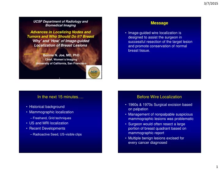

3/7/2015 UCSF Department of Radiology and Message Biomedical Imaging Advances in Localizing Nodes and • Image-guided wire localization is Tumors and Who Should Do It? Breast designed to assist the surgeon in ‘Why’ and ‘How’ of Image-guided successful resection of the target lesion Localization of Breast Lesions and promote conservation of normal breast tissue. Bonnie N. Joe, MD, PhD Chief, Women’s Imaging University of California, San Francisco In the next 15 minutes…. Before Wire Localization • 1960s & 1970s Surgical excision based • Historical background on palpation • Mammographic localization • Management of nonpalpable suspicious – Freehand, Grid techniques mammographic lesions was problematic • US and MRI localization • Surgeon would often resect a large • Recent Developments portion of breast quadrant based on mammographic report – Radioactive Seed, US-visible clips • Multiple benign lesions excised for every cancer diagnosed 1
3/7/2015 Freehand Localization Freehand Localization • Iterative approximations to get close to target • Developed in late 1970s, early 1980s to • Risk of pneumothorax (needle pointing towards chest wall) maximize likelihood of including the questionable mammographic lesion in the resected tissue • Freehand insertion of a needle into the region of the suspicious lesion preoperatively Stephenson, American Journal of Roentgenology. 1980;135: 184-186. http://www.ajronline.org/doi/abs/10.2214/ajr.135.1.184 Early Grid Localization Early Grid Localization • One to two needles placed into grid openings closest to target (arrow) based on mammographic image • Developed in 1980s • Needle insertion parallel to chest wall – Reduce risk of pneumothorax • Difficult to judge depth of needle placement in orthogonal view – Two needles placed a different depths From Goldberg, et al. Radiology 1983:146(3):833-839 2
3/7/2015 Modern Mammographic Grid Mammographic Wire Localization Localization • Grid technique allows accurate targeting of lesion Pt imaged in • Orthogonal imaging allows adjustment open grid of needle to proper depth of target • Wire deployed after optimizing needle Clip marking position site of cancer Mammographic Wire Localization Mammographic Wire Localization “E-12” localization Needle inserted perpendicular to skin at site of clip 3
3/7/2015 Mammographic Wire Localization Mammographic Wire Localization Wire deployed with clip located at distal stiffener (circle) Orthogonal view to BB markers placed on determine needle nipple and at skin entry placement relative to clip site for reference US Localization Specimen Radiograph • For mammographically occult lesions – No marker clip; significantly migrated clip • Real-time visualization of needle Confirms removal placement through target of intact hook wire and clip • Post-procedure *noncompressed* mammogram – Minimize wire migration 4
3/7/2015 Breast MR Localization 3D visualization Breast MR Localization • For lesions not amenable to mammographic or sonographic localization • Prone position • Obligate lateral or medial approach • Uses grid compression device and open coil • MR-safe needles and wires Breast MR Wire Localization Radioactive Seed Localization Planning Verification Post Mammogram • Placement of I125 Seed prior to surgery seen on a • Intra-operative localization with Gamma different slice foci of susceptibility probe corresponding to 3 needle locations • Reports of equivalency to wire localized excision • May be performed days in advance Sagittal T1 Post-gad Sagittal T1 Verification Post-Procedure Mammogram Chiu JC, et al.Am Surg. 2014 Jul;80(7):675-9. Sung, et al. Eur J Radiol. 2013 Sep;82(9):1453-7. Giesbrandt, McDonough. Pract Radiat Oncol. 2013 Apr-Jun;3(2 Suppl 1):S20. 5
3/7/2015 Radioactive Seed Localization Radioactive Seed Localization • Process of handling radioactivity Seed deployment Specimen Radiograph nontrivial (strict CA regulations) via 18g needle • Seed must be tracked from birth to grave Seed – Multiple departments (rad onc, mammography, OR, pathology) Biopsy clip marker – Patient ID bracelet and tracking outside of facility – Protocol for lost or transected seeds Sung, et al. Eur J Radiol. 2013 Sep;82(9):1453-7. US Visible Clips US Visible Clips • Can be visualized intraoperatively by Metallic component Echogenic collagen visible radiographically visible by ultrasound US to assist localization • No radiation • Requires additional surgeon/OR time to localize clip under US • Reports of “lost” clips in specimen • US visibility can vary Eby, et al. Acad Radiol. 2010 Mar;17(3):340-7. Corsi et al, Am J Surg. 2014 Oct 12. pii: S0002-9610(14)00506-6. Corsi et al, Am J Surg. 2014 Oct 12. pii: S0002-9610(14)00506-6. http://www.sciencedirect.com/science/article/pii/S0002961014005066# 6
3/7/2015 Message • Image-guided wire localization is designed to assist the surgeon in successful resection of the target lesion Thank you for your attention and promote conservation of normal breast tissue. Bonnie.joe@ucsf.edu 7
Recommend
More recommend