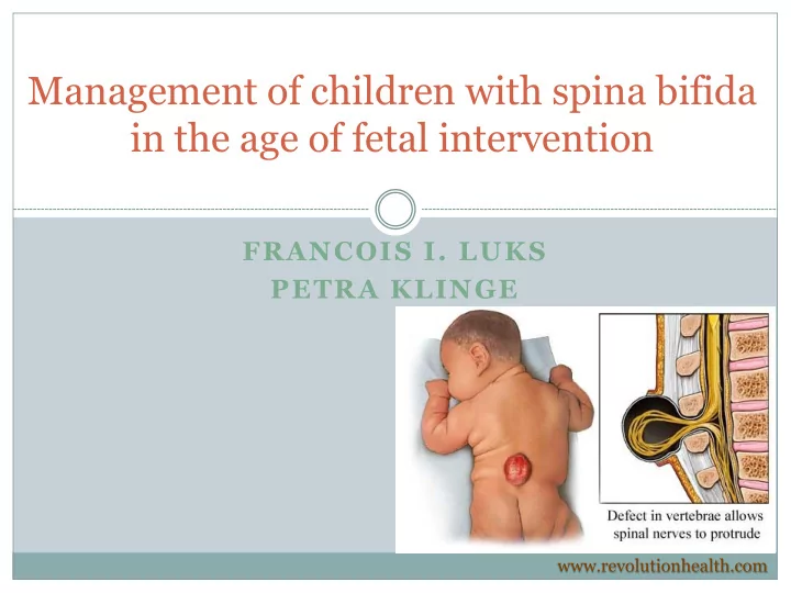

Management of children with spina bifida in the age of fetal intervention FRANCOIS I. LUKS PETRA KLINGE www.revolutionhealth.com
Spina Bifida and Neural Tube Defects Epidemiology One of the most common birth defects: 1-2 cases/1,000 births Certain populations have a greater risk: Highest incidence in Ireland and Wales More common in girls U.S.: 0.7/1,000 live births Higher on the East Coast than on the West Coast Higher in whites (1/1,000 births) Lower in African-Americans (0.1-0.4/1,000 births)
Spina Bifida and Neural Tube Defects Epidemiology Risk factors: Race and ethnicity Family history of neural tube defects Folate deficiency Medication/teratogenic effect: valproic acid Maternal age Diabetes Obesity Increased body temperature Hol FA et al, Clinical Genetics, 2008
Management of children with spina bifida in the age of fetal intervention Embryology of spina bifida Weeks 3-4 of gestation 3 phases: Neurulation Canalization Retrogressive differentiation
Spina Bifida and Neural Tube Defects Definitions and Classification Open spina bifida (Aperta) Meningocele in 5% Myelomeningocele (cord and cauda equina exposed) in 95% Closed spina bifida (Occulta) 50% have cutaneous stigmata Lipomyelomeningocele Filum terminale lipoma “Fatty” filum terminale Dermoid sinus and dermoid tumor
Spina Bifida and Neural Tube Defects Current management of spina bifida Primary treatment Perinatal care (protection of the neural tube, infections) Closure of the defect Management of hydrocephalus Chiari II hindbrain herniation Formal evaluation of spina bifida (overlaps with treatment) Physical examination: deformities, neuro exam; continence/tone Ultrasound MRI – brain MRI – spine Other: genetic testing, specialized imaging
Spina Bifida and Neural Tube Defects Definitive repair of the open neural tube defect Posterior vertebral defect Thecal sac Cord extruded into the sac (placode) Plate of embryonic epithelial cells: spinal cord
Spina Bifida and Neural Tube Defects Definitive repair of the open neural tube defect Closure within 24 hours No evidence that immediate/urgent closure improves function But: early closure reduces risk of infection Wound colonization after 36 hours Surgical technique: (neurosurgeon + plastic surgeon team) Placode dissected off arachnoid Allowed to drop into spinal canal Dura dissected off skin and lumbodorsal fascia Meninges Dura closed SKIN Placode CSF Muscular fascia closed FASCIA Skin closed
Spina Bifida and Neural Tube Defects Definitive repair of the open neural tube defect Surgical technique: Sharp microdissection of the placode
Spina Bifida and Neural Tube Defects Definitive repair of the open neural tube defect Continued dissection toward the placode Detethering Klinge, Taylor and Sullivan
Spina Bifida and Neural Tube Defects Definitive repair of the open neural tube defect Detethering of aberrant nerve roots Klinge, Taylor and Sullivan
Spina Bifida and Neural Tube Defects Definitive repair of the open neural tube defect Paraspinal muscle closure Klinge, Taylor and Sullivan
Spina Bifida and Neural Tube Defects Definitive repair of the open neural tube defect Klinge, Taylor and Sullivan
Spina Bifida and Neural Tube Defects Pathophysiology and associated disorders Hydrocephalus 80-95% incidence in myelomeningocele 100% of 35 thoracic lesions 88% of 114 lumbar lesions 68% of 40 sacral lesions Significant in 20% at birth Rintoul et al, Pediatrics 2002
Spina Bifida and Neural Tube Defects Management of hydrocephalus Imaging: ventriculomegaly (Ventricular index >0.33) Pediatric characteristics: Selective thinning of the occipiatl cranial vault and cortex: Rigid nuclear masses (basal ganglia) in the frontal lobe Monitor head circumference! Ventricular index > 0.33 47.65 mm 137.96 mm
Spina Bifida and Neural Tube Defects Management of hydrocephalus Serial head ultrasounds in the newborn:
Spina Bifida and Neural Tube Defects Management of hydrocephalus Temporary drainage: Lumbar puncture External ventricular drainage, reservoir Shunt Weight >2.5 kg No active infection Medically stable Endoscopic third ventriculostomy
Spina Bifida and Neural Tube Defects Management of hydrocephalus Types of shunts: Adjustable valves
Spina Bifida and Neural Tube Defects Management of hydrocephalus Endoscopic third ventriculostomy
Spina Bifida and Neural Tube Defects Pathophysiology and associated disorders Chiari II malformation 99% of myelomeningocele have radiographic Chiari II Only symptomatic ones require treatment (30% at 5 years) Responsible for 15-20% of deaths in children with MMC Respiratory failure/arrest Syringomyelia
Spina Bifida and Neural Tube Defects Treatment of Chiari II malformation
Spina Bifida and Neural Tube Defects Current management of spina bifida Secondary management Relatively recent: now that these children survive long-term The most difficult – chronic vigilance CNS monitoring: VP shunt management Management of tethered cord (10%) Physical therapy evaluation/motor function of lower extremities Preventive medicine – insensate lower body Psychological support
Spina Bifida and Neural Tube Defects Current management of spina bifida Secondary management Management of tethered cord Second detethering surgery for decline in function and/or before correction of scoliosis after surgery Tethering at the MMC closure site
Spina Bifida and Neural Tube Defects Which organ systems does it affect? Neuro-motor Neurodevelopmental, hydrocephalus, CNS development
Spina Bifida and Neural Tube Defects Which organ systems does it affect? Neuro-motor Neurodevelopmental, hydrocephalus, CNS development Urogenital Gastrointestinal Gastroesophageal reflux disease (GERD) Constipation More commonly: incontinence Other Variability in severity for all systems (GI specifically)
Management of children with spina bifida Spina Bifida and Neural Tube Defects in the age of fetal intervention Peripheral effects of open neural tube defect Exposed spinal cord during gestation (Progressive?) damage to the exposed neural tube Variable paresis, urine & stool incontinence CSF leak into amniotic cavity Basis for prenatal testing: leakage of alpha-fetoprotein (AFP) Increased concentration in the amniotic fluid (amniocentesis) Maternal Serum AFP (MSAFP) elevated as well False-positives: any other cause of AFP leakage: gastroschisis
Management of children with spina bifida Spina Bifida and Neural Tube Defects in the age of fetal intervention Peripheral effects of open neural tube defect Exposed spinal cord during gestation (Progressive?) damage to the exposed neural tube Could spina bifida be cured – or even prevented ?
Management of children with spina bifida in the age of fetal intervention Embryology of spina bifida – can it be prevented? Progressive development theory Is only one theory – and the most simplistic one Prolonged in utero exposure of the neural tube leads to Chronic leakage of CSF Gradual siphoning and hindbrain herniation Increased risk of hydrocephalus Progressive damage to the neural placode Progressive peripheral nerve damage • Lower extremity function • Sphincter function
Management of children with spina bifida in the age of fetal intervention Spina bifida – can it be diagnosed in utero? Ultrasound Spinal defect “Lemon” sign: abnormally shaped skull (head circumference) “Banana” sign: abnormally shaped cerebellum Hydrocephalus
Management of children with spina bifida in the age of fetal intervention Spina bifida – can it be diagnosed in utero? Magnetic Resonance Imaging
Management of children with spina bifida in the age of fetal intervention Animal experiments – Fetal sheep Creation of a neural tube defect in a mid-gestation lamb: Leads to phenotype resembling clinical spina bifida Causes hind limb paralysis Causes hydrocephalus Normal Spina bifida Repaired Spina bifida Meuli M et al, Nature Medicine 1995
Management of children with spina bifida in the age of fetal intervention Animal experiments – Fetal sheep Creation of a neural tube defect in a mid-gestation lamb: Leads to phenotype resembling clinical spina bifida Causes hind limb paralysis Causes hydrocephalus Closure of the defect in utero: Corrects all these problems Meuli M et al, Nature Medicine 1995
Management of children with spina bifida in the age of fetal intervention Animal experiments – Fetal sheep Creation of a neural tube defect in a mid-gestation lamb: Leads to phenotype resembling clinical spina bifida Causes hind limb paralysis Causes hydrocephalus Closure of the defect in utero: Corrects all these problems Caveat: because this is a surgical created, then corrected defect, it may not be the same as the clinical syndrome
Recommend
More recommend