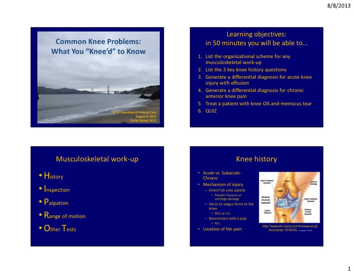

8/8/2013 Learning objectives: Common Knee Problems: in 50 minutes you will be able to… What You “Knee’d” to Know 1. List the organizational scheme for any musculoskeletal work-up 2. List the 3 key knee history questions 3. Generate a differential diagnosis for acute knee injury with effusion 4. Generate a differential diagnosis for chronic anterior knee pain 5. Treat a patient with knee OA and meniscus tear 6. QUIZ UCSF Essentials of Primary Care August 8, 2013 Carlin Senter, M.D. Musculoskeletal work-up Knee history • Acute vs. Subacute- • H istory Chronic • Mechanism of injury • I nspection – Direct fall onto patella • Patellar fracture or • P alpation cartilage damage – Varus or valgus force to the knee • R ange of motion • MCL or LCL – Noncontact with a pop • ACL • O ther T ests http://www.ski-injury.com/kneeanat.gif, • Location of the pain Accessed 10/04/05. Accessed 10/4/05 1
8/8/2013 3 key knee injury questions Case #1: House of Air • 35 y/o woman on trampoline half-pipe. 1. Locking = meniscus or intra-articular loose body Jumped down and felt a pop with immediate knee pain and swelling. 2. Instability = ligament • Went to ER: placed in knee immobilizer and 3. Swelling = intra-articular derangement given Vicodin for pain relief. 1. Immediate: due to blood (ACL, fracture, patellar • Now, 3d later, has posterior pain and tightness dislocation) 2. Subacute: 8-24 hours, due to synovial with bending. • Knee feels unstable if not in the brace. inflammation (meniscus, MCL) Ddx acute traumatic knee injury with Musculoskeletal exam order effusion • H istory • Intra-articular derangement • I nspection – (+) instability � ligament • P alpation – (+) locking � meniscus – Dislocation • R OM • Patella • Knee • O ther – Cartilage damage – Patellar or quad tendon • T ests rupture http://www.ski-injury.com/kneeanat.gif, Accessed 10/04/05. Accessed 10/4/05 2
8/8/2013 Knee exam case #1: Inspection Significance of acute effusion • Intra-articular derangement • You will likely be ordering xray +/- MRI • The patient will not be returning to sport today Knee exam case #1: Palpation Palpation: patellar facet Ballottement Video courtesy of Dr. Anthony Luke 3
8/8/2013 Knee exam case #1: Palpation Knee exam case #1 • Supine, knee fully extended • ROM: 5-90, limited due – Ballotement to evaluate for effusion to pain (normal 0-135) – Medial patellar facet (patellar dislocation) – Determine if knee is – Patellar apprehension (patellar dislocation) • Straight leg raise intact locking or if ROM is – If not - Quad tendon or patellar tendon rupture -> urgent ortho limited due to effusion • Knee flexed to 90 degrees – Locking: think bucket – Joint line (meniscus) handle meniscus. – Lateral femoral condyle (patellar dislocation) • Urgent xrays, MRI – Above and below medial and lateral joint lines (MCL, LCL) • Urgent referral to sports • Our patient: tender medial joint line, can do straight leg surgeon for arthroscopy raise – Rules out patellar dislocation, LCL, tendon rupture Knee exam case #1 Other Tests: Lachman to evaluate ACL • Strength 5/5 hip flexion, knee extension, PF, DF. – (+) active knee extension rules out quad or patellar tendon rupture • 2+ dorsalis pedis pulses bilaterally • Sensation intact to light touch over legs bilaterally • Reflexes 2+ at patella and achilles bilaterally Video courtesy of Dr. Anthony Luke 4
8/8/2013 PCL: Sag sign PCL: Posterior Drawer Video courtesy of Dr. Anthony Luke MCL and LCL Meniscus: McMurray Sensitivity medial 65%, Specificity medial 93% Video courtesy of Dr. Anthony Luke Magee, DJ. Orthopaedic Physical Assessment, 5 th ed. 2008. 5
8/8/2013 Meniscus: Thessaly Meniscus: squat Video courtesy of Dr. Anthony Luke Case #1 special tests Case #1 diagnosis A. Patellar tendon rupture • (+) pain with medial McMurray, (-) lateral B. PCL tear • (+) Thessaly – medial pain C. ACL tear • (+) Squat – medial pain D. MCL tear • (-) laxity to varus or valgus at 0 and 30 E. Meniscus tear F. ACL + meniscus tear • (+) Lachman without endpoint 78% • (-) Posterior drawer http://www.ski-injury.com/kneeanat.gif, 20% Accessed 10/04/05. Accessed 10/4/05 2% 0% 0% 0% o... us... r ar L tear us tear tea r tend CL te isc CL C n M isc e P A CL + m Patella n e M A 6
8/8/2013 Traumatic knee effusion red flags � Case #1 treatment urgent ortho referral • Knee brace +/- crutches • Locked knee: unable to fully extend compared depending on pain and to other side instability • Xrays to r/o fracture – Bucket handle meniscus • MRI to confirm diagnosis – Make non weight bearing w/crutches • Pain medication • Fracture (tibial plateau, patella) • PT to restore normal ROM, decrease swelling, • Unable to extend knee against gravity strengthen quad • Orthopaedic surgery – Patellar or quadriceps tendon rupture referral to discuss +/- – Needs urgent surgical repair reconstruction Segond fracture – avulsion of lateral tibial plateau in ACL tear Case #2: Sketcher Shape-Ups Subacute knee history • 3 key questions 40 y/o woman with sharp anterior knee pain x 1 – Swelling month. Might have some swelling. No locking – Locking but the knee is popping. Feels unstable when – Instability walking down stairs. Pain worse up/down stairs. Painful when gets up from sitting. Exercise: • Exercise and activity history: squats, lunges, started a walking program for New Year’s new training program, marathon? resolution, wearing new Sketcher Shape-Up • Shoes: how old, what type shoes. No squats/lunges. Doesn’t wear • Orthotics: how old, why wearing them orthotics. 7
8/8/2013 Ddx subacute-chronic anterior knee Case #2: Inspection pain 1. Patellofemoral pain syndrome 2. Patellar chondromalacia 3. Osteochondral lesion 4. Osteoarthritis of patellofemoral joint Patellofemoral pain syndrome: Case #2: Palpation miserable malalignment syndrome • Effusion: none • Femoral anteversion • Joint line, patellar facets (inward rotation of – Tender medial and femur) • Squinting patella lateral patellar facets (inward patellar rotation) • Patella alta • Increased Q-angle • Excessive outward tibial http://www.kneeguru.co.uk/KNEEnotes/node/763 rotation http://www.gla.ac.uk/ibls/US/fab/tutorial/biomech/akp3.html 8
8/8/2013 Case #2: ROM Case #2: Other tests • 0-135 • Ligaments: no laxity – Lachman • (+) crepitus with flexion and extension as – Posterior drawer patella moves across articular surface of femur – MCL – LCL • Meniscus: no pain – McMurray Case #2: Other tests Ober part 1 identify tightness and weakness • Ober (too tight?) • Hip abduction strength (weak?) • One-legged standing squat (weak? Pain?) Passive hip abduction and extension. Hip extension � ITB positioned over greater trochanter of femur. http://www.youtube.com/watch?v=A0C0WBw4l4s&feature=player_detailpage 9
8/8/2013 Ober part 2 Hip abduction strength Lower the upper limb. If tight ITB then hip will not adduct past neutral. Compare side to side. http://www.youtube.com/watch?v=9Iy-QrcuGno&feature=player_detailpage http://www.youtube.com/watch?v=A0C0WBw4l4s&feature=player_detailpage One-legged standing squat One-legged standing squat • Patient standing on unaffected leg • Do 3 slow 1-legged squats • Watch for stability, valgus angulation of knee, ask about pain • Switch and perform on affected leg • Sign of weak hip abductors, weak core • Can bring out pain of patellofemoral pain 10
8/8/2013 Case #2: Sketcher Shape-Ups One-legged standing squat Physical exam • Valgus angulation of the knees • No effusion • Tender medial and lateral patellar facets • ROM 0-135, crepitus • No laxity with lachman, posterior drawer, varus or valgus at 0 and 30 degrees • (+) Ober bilaterally • 4/5 hip abductor strength bilaterally • Unstable 1-legged squat with valgus knee angulation Case #2 diagnosis Case #2 treatment • Physical therapy rx A. Patellofemoral pain syndrome – Strengthen hip abductors 89% B. Patellar chondromalacia – Strengthen quadriceps – Stretch ITB, quads, hamstrings C. Osteochondral lesion • Correct alignment: consider OTC orthotics with D. Osteoarthritis arch support if pes planus • Activity: avoid running, squats, lunges, stair- running, downhill hiking until improved. • If not improved with above � xrays and if those 8% 3% 0% normal then MRI (or refer to sports medicine) . . . s . . . i . . . t a l d l i a r r n r h o o d t m h n r c a e o o f r h o a e c t l l l o l e s e e O t t a t a s P P O 11
Recommend
More recommend