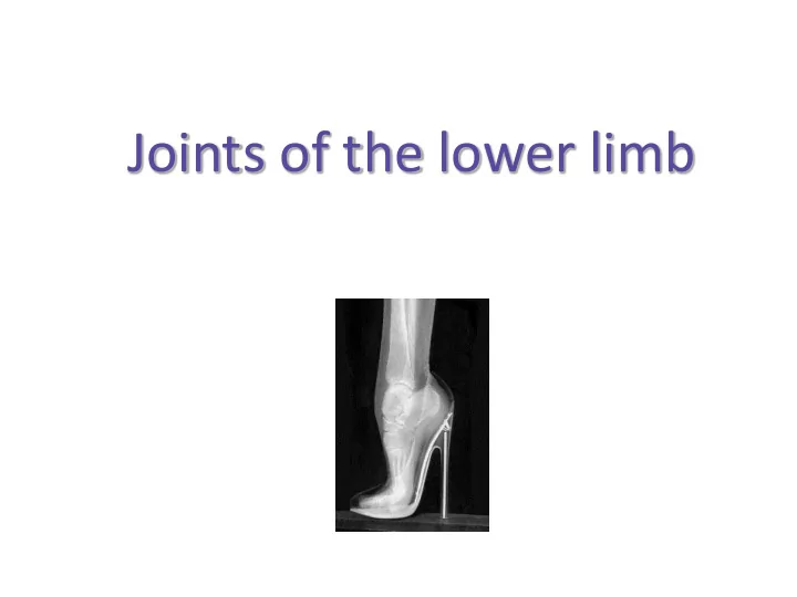

Joints of the lower limb
Joints of the lower limb Juncturae membri inferioris Joints of the pelvic girdle ( juncturae cinguli membri inferioris ) – articulatio sacroiliaca (sacro-iliac joint) – symphysis pubica (pubic symphysis) Joints of the freelower limb ( juncturae membri inferioris liberi ) – art. coxae (hip joint) – art. genus (knee joint) – art. talocruralis (ankle joint) – joints of foot
Joints of the pelvic girdle Juncturae cinguli membri inferioris • articulatio sacroiliaca (sacro-iliac joint) • synarthroses cinguli pelvici – symphysis pubica (pubic symphysis) – syndesmoses • membrana obturatoria • lig. sacrospinale et sacrotuberale • lig. inguinale ( Pouparti ) Čihák Radomír, Anatomie 1, Druhé, upravené a doplněné vydání, Grada Publishing 2001
Sacroiliac joint Articulatio sacroiliaca http://flickrhivemind.net/Tags/xraycost
Sacroiliac joint ( Articulatio sacroiliaca ) Type simple, 3-axial, plane, stiff (amphiarthrosis) Head facies auricularis ossis sacri (we do not distinguish head and cavity) Fossa facies auricularis ossis ilii (we do not distinguish head and cavity) Articular capsule and lig. sacroiliacum anterius, posterius et interosseus (between tuberositas iliaca et ligaments tuberositas ossis sacri), lig. iliolumbale minimal, ventrodorsal and nutation around S2, for example when walking and Movements jumping change of gravity center – change of pelvis position (pelvis inclination) Neutral position = basic position non-corresponding articular surfaces – reduction of movement extent with ageing – overload of back muscles – pain in lumbar vertebrae with irradiation Notes into the thigh
Ligaments of sacroiliac joint
CT of sacroiliac joint
CT pánve
Pubic symphysis ( Symphysis pubica ) • facies symphysialis ossis pubis bilaterally • discus interpubicus (width 4-5 mm ) – fibrous cartilage, hyaline at margins, inside may be a cavity • eminentia retropubica – protrudes dorsally into lesser pelvis – palpable per vaginam • lig. pubicum superius et inferius – inferior ligament very strong! • almost no movements – relaxes during gravidity due tohormone relaxine Čihák Radomír, Anatomie 1, Druhé, upravené a doplněné vydání, Grada Publishing 2001
Diastasis of pubic symphysis („open book “ fracture) 11 http://en.wikipedia.org/wiki/File:Diastasis_symphysis_pubis_1300500.JPG
Syndesmoses of pelvic girdle • ligamentum inguinale Pouparti (inguinal ligament) – between spina iliaca anterior superior and tuberculum pubicum – false ligament = caudal margin of aponeurosis (flat tendon) of musculus obliquus externus abdominis – under ligament topographic sites: lacuna vasorum et musculorum (passages for vessels and muscles) • ligamentum sacrospinale et sacrotuberale – stability of pelvis – form topographic sites: foramen ischiadicum majus et minus • membrana obturatoria – canalis obturatorius
Čihák Radomír, Anatomie 1, Druhé, upravené a doplněné vydání, Grada Publishing 2001
Pelvis as a whole • composed of paired os coxae and unpaired os sacrum • inclinatio pelvis (inclination) = 60 ° • pelvic circle – tough and elastic complex of bones and ligaments transferring weight of trunk on lower limbs • linea terminalis – circular line running via: promontorium ossis sacri linea arcuata eminetia ilopubica pecten ossis pubis superior margin of symphysis pubica – separates pelvis major and pelvis minor
Linea terminalis http://home.comcast.net/~wnor/pelvis.htm
Pelvis as a whole • pelvis major (greater pelvis) – small intestine and a part of large intestine • pelvis minor (lesser pelvis) – urinary bladder and urethra, prostate ♂ / ovarium, uterine tube, uterus, vagina ♀ and rectum • weight of the trunk tilts the upper part of os sacrum ventrally • movement axis runs horizontally via the articular surfaces of sacroiliac joint • lower part of os sacrum tilts dorsally and stabilizes the pelvis in ventrodorsal direction • lig. sacrospinale et sacrotuberale limit tilting movements of the lower part of os sacrum dorsally
Clinical notes Articulatio sacroiliaca (sacroiliac joint, SI joint) • very strong ligaments, only small tilting movements, very important for pelvis inclination change ( inclinatio pelvis ) when changing the gravity center • overload of SI joint (sport, work in bending position, arthrotic changes) leads to pain syndrome – local pain + irradiation into the thigh – result of the spasm (spasmic contraction) of surrounding muscles stabilizing the overloaded joint Symphysis pubica (pubic symphysis) • eminentia retropubica – decreases ventrodorsal diameter of pelvic aditus, palpable in vaginal (obstetric) examination • tissue of symphysis is during delivery relaxed by ovarian hormone relaxine – enlargement of space for fetus passage (and coccyx moves dorsally) • strenthened by ligaments – even in case of symphyseolysis the lig. pubicum inferius holds bones together
Joints of free lower limb Juncturae membri inferioris liberi • diarthroses – articulatio coxae s. coxofemoralis (hip joint) – articulatio genus (knee joint) – articulatio tibiofibularis – articulationes pedis • art. talocruralis (ankle joint) • art. subtalaris s. talocalcanea (subtalar joint) • art. tarsi transversa (Choparti) – art. talocalcaneonavicularis – art. calcaneocuboidea • art. cuneonavicularis • articulationes intercuneiformes • articulationes tarsometatarsales • articulationes intermetatarsales • articulationes metatarsophalangeae • articulationes interphalangeae pedis • synarthroses – syndesmosis tibiofibularis • membrana interossea cruris • lig. tibiofibulare anterius et posterius
Hip joint ( Articulatio coxae ) Type of joint simple, 3-axial, limited ball-and-socket (art. cotylica), mobile Head caput femoris Fossa acetabulum: facies lunata + labrum acetabuli capsule: ventrally as far as linea intertrochanterica, dorsally in 2/3 of collum femoris lig. iliofemorale ( Bigellowi ) – thickest ligament in human body , lig. Articular capsule ischiofemorale, lig. pubofemorale, zona orbicularis, lig. capitis femoris, lig. and its ligaments transversum acetabuli retinacula Weibrechti – blood supply to capit femoris (branches of a. circumflexa femoris medialis) labrum acetabuli, pulvinar acetabuli, lig. capitis femoris Special structures (false intra-articular ligament covered with synovial membrane) flexion-extension (=dorsal flexion), adduction-abduction, Movements (+hyperadduction), internal-external rotation, circumduction Neutral position mild flexion, slight abduction and external rotation lig. iliofemorale limits extension lig. pubofemorale limits abduction and external rotation lig. ischiofemorale limits adduction and internal rotation Notes CCD (capitocolodiaphyseal) angle = 126 ° developmental joint dysplasia (developmental dysplasia of hip joint) – most common defect of locomotion system
Ligaments of hip joint
http://pds.exblog.jp/pds/1/200511/02/86/a0057586_14531191.jpg
http://www.centenoschultz.com/wp-content/images/ishiofemoral-liagment.jpg
Ligamentum capitis femoris
Ligaments of hip joint • lig. capitis femoris • zona orbicularis – lig. ischiofemorale – lig. pubofemorale • lig. transversum acetabuli http://www.bartleby.com/107/Images/small/image341.jpg http://upload.wikimedia.org/wikipedia/commons/thumb/6/65/Gray343.png/250px-Gray343.png
http://childrenshospital.org/clinicalservices/Site1163/Images/Hip.jpg
Movements in hip joint • flexion up to 120 ° • extension up to 13 ° • abduction up to 40 ° • adduction up to 10 ° • rotation – external up to 15 ° – internal up to 35 °
Clinical notes • transfer of great forces is done by architecture of acetabulum, os ilium and proximal femur (acetabular pillar, Ward‘s triangle, Adams‘ arch ) • hip joint luxation occurs mostly in high-energy injuries – typical is hit of the car dash board („dash - board“ injury) • in 90% cases it is the posterior luxation (into weak spots between ligaments) • 10% anterior luxation – rare obturator luxation
Structures inside femur • Adams‘ arch – onn medial side; collum femoris continues into the diaphysis by thick cortical bone • Ward‘s triangle – space inside the collum femoris between band of crossing trabecules is a cavity filled with yellow bone marrow Ward‘s triangle Adams‘s arch
Hip joint replacement • TEP – total endoprosthesis • CEP – cervicocapital endoprosthesis • one of the most common operations in orthopaedics • arthrotic changes in joint, fractures of collum, fractures of acetabulum
Recommend
More recommend