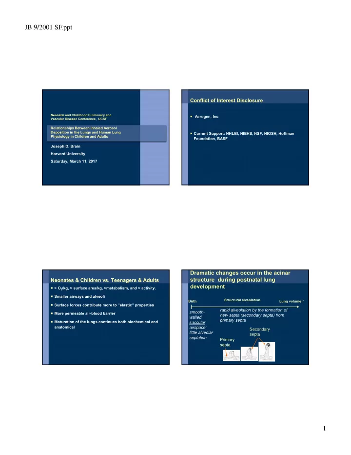

JB 9/2001 SF.ppt Conflict of Interest Disclosure Neonatal and Childhood Pulmonary and Aerogen, Inc Vascular Disease Conference , UCSF Relationships Between Inhaled Aerosol Deposition in the Lungs and Human Lung Current Support: NHLBI, NIEHS, NSF, NIOSH, Hoffman Physiology in Children and Adults Foundation, BASF Joseph D. Brain Harvard University Saturday, March 11, 2017 Dramatic changes occur in the acinar structure during postnatal lung Neonates & Children vs. Teenagers & Adults development > O 2 /kg, > surface area/kg, >metabolism, and > activity. Smaller airways and alveoli Structural alveolation Birth Lung volume ↑ Surface forces contribute more to ”elastic” properties rapid alveolation by the formation of smooth- More permeable air-blood barrier new septa (secondary septa) from walled primary septa Maturation of the lungs continues both biochemical and saccular anatomical airspace; Secondary little alveolar septa septation Primary septa 1
JB 9/2001 SF.ppt human birth 1.5~2y ~8years adult rat b. 4d 7d 14d 21d 35d > 90 days Semmler-Behnke et al. , PNAS 2012 What is an Aerosol? Aerosols A suspension of small solid or liquid particles in a gas (usually air) Particle size determines how aerosols move through air and Unit of size measurement for respirable aerosols is the where and how they interact with the surfaces they encounter. micrometer (µm) = micron (µ) = 10 -6 m Breathing pattern and lung anatomy also determine deposition of aerosols in the respiratory tract. Size ranges: Coarse 2.5–10.0 µm Fine < 2.5 µm Ultrafine < 0.1 µm Environmental and pharmacologic aerosols are composed of a range of particle sizes. Monodisperse Same size – unusual Polydisperse A continuous spectrum of sizes 2
JB 9/2001 SF.ppt Aerosols: Are ubiquitous indoors and outdoors Cause and aggravate lung disease Can be an important therapeutic tool Aerosols Can Hurt Neonates and Children Aerosols Can Heal Neonates and Children Particulate Matter – Indoors, Outdoors, Occupational Pulmonary Surfactants Mold, E.G. Stachybotrys chartarum Antivirals, Antibiotics, e.g. Tobramycin Passive Tobacco Smoke Steroids Airborne Infection: Measles, Tb, Influenza Bronchodilators Mucolytic, e.g. Pulmozyme Neonate and child lungs not only collect toxic particles, they Anti-proteases, inhibitors of fibrosis or abnormal repair also capture therapeutic aerosols 3
JB 9/2001 SF.ppt Drug Delivery Directly to the Lungs Routes of Exposure and Anatomic Barriers Rapid delivery to the site of action; the lungs are < 1% of body weight, why waste it. Route Organ Barrier Surface Area Thickness from Typical Daily (m 2 ) Environment to Exposure High local concentrations where the drug is needed Blood ( m) Dermal Skin Epidermis 1.8 100-1000 Variable, Minimize dosing of extra-pulmonary sites and thus easily reduced minimize side effects Oral GI Intestinal 1,200 15-40 1.5 kg food Ingestion Tract Epithelium 2 kg water Some particles from Avoids hepatic uptake and processing. respiratory tract 10-20 m 3 Inhalation Lungs Alveolar 140 0.64 Injections, IV administration, not needed. Moreover if Epithelium (10,000 – 20,000 L) or 15 - 25 kg air the drug is needed on alveolar suraces, the tight Two notable characteristics of the lung: epithelial is avoided • high daily exposure • thin barrier Drug Delivery Through the Lungs Is Possible Advantages of pulmonary delivery of proteins to systemic circulation: Large surface area—about 150 m 2 Thin barrier—< 1 µm Few proteases Avoids first-pass liver clearance Rapid and efficient transport Avoids unpleasant injections 4
JB 9/2001 SF.ppt Even Peptide and Protein Hormones Can Be Administered Via the Lungs Insulin Somatostatin Calcitonin Thyroid stimulating hormone Parathyroid hormone α -1-antitrypsin Human growth hormone Niven RW. Crit Rev Ther Drug Carrier Syst . 1995;12:151-231. Wolff RK, Dorato MA. Crit Rev Toxicol . 1993;23:343-369. Pharmacologic Aerosols: Deposition of Particles The dose of an inhaled aerosolized drug and its anatomic distribution depend on: aerosol size & concentration, physics breathing pattern (IMPACTION) lung anatomy, e.g. acute and chronic changes (asthma, ARDS, PAH) equipment between the device producing the aerosol and the patient’s airway 5
JB 9/2001 SF.ppt Aerodynamic particle diameter is a determinant of how much and Classification of Particles by Size where particles are deposited in the respiratory tract Inhalable Coarse Particles, PM >10 m • Can deposit in the nose or mouth (IMPACTION) • Deposited primarily by impaction Thoracic Coarse Particles, PM 2.5 to 10 m • Can deposit in airways of the lungs • Deposited primarily by impaction and sedimentation Respirable Particles (fine fraction) PM <2.5 m • Most penetrate beyond the terminal bronchioles and reach the gas exchange region • Deposit there primarily by sedimentation and diffusion Primary mechanism Inertial Impaction Ultrafine Particles are <0.1 m (also called nanoparticles) of deposition Diffusion Sedimentation • Small fraction by mass but not by number or surface area. • Deposit primarily by diffusion throughout the respiratory tract Now let us review: Effect of Breathing Pattern on Particle Deposition Increased linear velocity of airflow promotes inertial Lung anatomy impaction of particles in more proximal airways. When inspiratory flow rates are excessive, there may be Clearance mechanisms that determine significant losses in the device and oral phatynx. drug retention An increase in tidal volume leads to deeper penetration by particles, more alveolar deposition. Lower inspiratory flow rates and increased end tidal breath holds provide more time for diffusion and sedimentation and thus more alveolar deposition. 23 6
JB 9/2001 SF.ppt CONDUCTING AIRWAYS NASOPHARYNX (upper airways) Each clump of alveoli & ducts is called an acinus. Air is filtered, humidified and brought to body Gas exchange occurs here. Temperature in the conducting airways. 7
JB 9/2001 SF.ppt Rates of Particle Clearance Approximate Anatomic Clearance Clearance Half- Region Mechanism time Nasopharynx Mucociliary Minutes Transport Tracheobronchial Mucociliary Minutes to Hours Transport Alveolar Macrophage Hours Phagocytosis Alveolar Macrophage Hours to Months to Dissolution ? Alveolar Macrophage 8
JB 9/2001 SF.ppt Iron Oxide Particles on Respiratory Surfaces of the Lung 3 hours after a 1 hour exposure Arrows indicate macrophages that have taken up iron oxide particles. Austin et al. AJRCCM 195(1):23-31, 2017 Feb. 1 Conclusions: Give neonates and children clean air. Take advantage of therapeutic aerosols Modify devices and delivery strategies as needed. Early events have long term, life time consequences. Austin et al. AJRCCM 195(1):23-31, 2017 Feb. 1 9
Recommend
More recommend