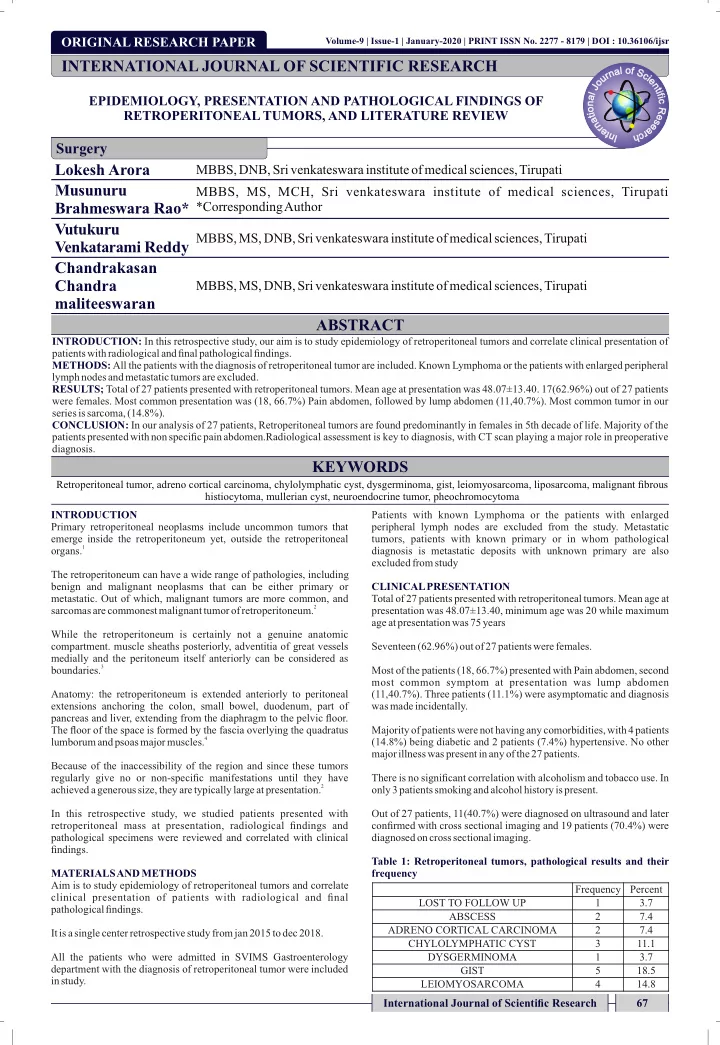

ORIGINAL RESEARCH PAPER age at presentation was 75 years Out of 27 patients, 11(40.7%) were diagnosed on ultrasound and later only 3 patients smoking and alcohol history is present. There is no significant correlation with alcoholism and tobacco use. In major illness was present in any of the 27 patients. (14.8%) being diabetic and 2 patients (7.4%) hypertensive. No other Majority of patients were not having any comorbidities, with 4 patients was madeincidentally. (11,40.7%). Three patients (11.1%) were asymptomatic and diagnosis most common symptom at presentation was lump abdomen Most of the patients (18, 66.7%) presented with Pain abdomen, second Seventeen (62.96%) out of 27 patients were females. presentation was 48.07±13.40, minimum age was 20 while maximum diagnosed on cross sectional imaging. Total of 27 patients presented with retroperitoneal tumors. Mean age at CLINICAL PRESENTATION excluded from study diagnosis is metastatic deposits with unknown primary are also tumors, patients with known primary or in whom pathological peripheral lymph nodes are excluded from the study. Metastatic Patients with known Lymphoma or the patients with enlarged in study. department with the diagnosis of retroperitoneal tumor were included All the patients who were admitted in SVIMS Gastroenterology It is a single center retrospective study from jan 2015 to dec 2018. pathological findings. confirmed with cross sectional imaging and 19 patients (70.4%) were Table 1: Retroperitoneal tumors, pathological results and their Aim is to study epidemiology of retroperitoneal tumors and correlate 2 4 LEIOMYOSARCOMA 18.5 5 GIST 3.7 1 DYSGERMINOMA 11.1 3 CHYLOLYMPHATIC CYST 7.4 ADRENO CORTICAL CARCINOMA frequency 7.4 2 ABSCESS 3.7 1 LOST TO FOLLOW UP Percent Frequency Volume-9 | Issue-1 | January-2020 | PRINT ISSN No. 2277 - 8179 | DOI : 10.36106/ijsr 67 International Journal of Scientific Research Surgery INTERNATIONAL JOURNAL OF SCIENTIFIC RESEARCH clinical presentation of patients with radiological and final AND METHODS EPIDEMIOLOGY, PRESENTATION AND PATHOLOGICAL FINDINGS OF ABSTRACT KEYWORDS diagnosis. Radiological assessment is key to diagnosis, with CT scan playing a major role in preoperative patients presented with non specific pain abdomen. CONCLUSION: In our analysis of 27 patients, Retroperitoneal tumors are found predominantly in females in 5th decade of life. Majority of the series is sarcoma, (14.8%). were females. Most common presentation was (18, 66.7%) Pain abdomen, followed by lump abdomen (11,40.7%). Most common tumor in our RESULTS; Total of 27 patients presented with retroperitoneal tumors. Mean age at presentation was 48.07±13.40. 17(62.96%) out of 27 patients lymph nodes and metastatic tumors are excluded. METHODS: All the patients with the diagnosis of retroperitoneal tumor are included. Known Lymphoma or the patients with enlarged peripheral patients with radiological and final pathological findings. INTRODUCTION: In this retrospective study, our aim is to study epidemiology of retroperitoneal tumors and correlate clinical presentation of MBBS, MS, DNB, Sri venkateswara institute of medical sciences, Tirupati histiocytoma, mullerian cyst, neuroendocrine tumor, pheochromocytoma maliteeswaran Chandra Chandrakasan Vutukuru Author *Corresponding MBBS, MS, MCH, Sri venkateswara institute of medical sciences, Tirupati Brahmeswara Rao* Musunuru MBBS, DNB, Sri venkateswara institute of medical sciences, Tirupati Lokesh Arora RETROPERITONEAL TUMORS, AND LITERATURE REVIEW Retroperitoneal tumor, adreno cortical carcinoma, chylolymphatic cyst, dysgerminoma, gist, leiomyosarcoma, liposarcoma, malignant fibrous INTRODUCTION MATERIALS extensions anchoring the colon, small bowel, duodenum, part of findings. pathological specimens were reviewed and correlated with clinical retroperitoneal mass at presentation, radiological findings and In this retrospective study, we studied patients presented with achieved a generous size, they are typically large at presentation. 2 regularly give no or non-specific manifestations until they have Because of the inaccessibility of the region and since these tumors lumborum and psoas major muscles. 4 The floor of the space is formed by the fascia overlying the quadratus pancreas and liver, extending from the diaphragm to the pelvic floor. Anatomy: the retroperitoneum is extended anteriorly to peritoneal Primary retroperitoneal neoplasms include uncommon tumors that boundaries. 3 medially and the peritoneum itself anteriorly can be considered as compartment. muscle sheaths posteriorly, adventitia of great vessels While the retroperitoneum is certainly not a genuine anatomic sarcomas are commonest malignant tumor of retroperitoneum. 2 metastatic. Out of which, malignant tumors are more common, and benign and malignant neoplasms that can be either primary or The retroperitoneum can have a wide range of pathologies, including organs. 1 emerge inside the retroperitoneum yet, outside the retroperitoneal 14.8 Venkatarami Reddy MBBS, MS, DNB, Sri venkateswara institute of medical sciences, Tirupati
Recommend
More recommend