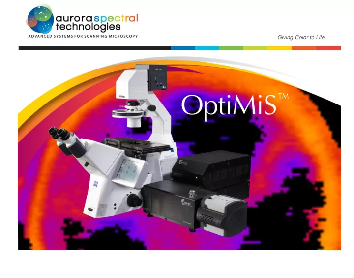

Giving Color to Life
W i d e f i e l d, C o n f o c a l a n d Two-Photon Microscop y Systems with Exquisite Spectral Resolution Aurora Spectral Technologies is pleased to offer state-of-the-art optical microscopy OptiMiS is the only scanning system that permits real-time quantitative analysis of and micro-spectroscopy (OptiMiS) technology capable of generating unparalleled single molecules and molecular complexes and their spatial distribution in living data for biological and biophysical research. AST products and services range from cells. The system is capable of producing a full set (up to 200 wavelength channels standard confocal and multiphoton (two-photon) microscopes and accessories to over 425-650 nm) of spectrally resolved images with a single excitation scan. In our powerful optical micro-spectroscopy technology, OptiMiS TruLine. The addition, it features sensitivity much higher (by over 100×) than any point-scan OptiMiS can be attached to the customer’s microscope or sold as a complete confocal or two-photon (multiphoton) microscope in the market. system. O P T I MI S I N T E G R AT I ON P A CK A GE This package includes all of the components required to configure our syst e m with a laser, microscope, and camera. It comprises a periscope system to a lig n the laser beam, a computer controlled laser power modulator, and all o f the elements needed for complete assembly of the system. I m po rt a n tly , t h e package a l s o i nc l ud e s pr e c i s i on alignment by a s k ill e d i n st rum e nt s p e c i a li st and two days of O P T IM I S S Y S TE M h a nd s -on training. This is our proprietary product, which may be attached to existing r e s ea r ch ANDOR iXON ™ EMCCD CA M E R A m i cro s cop e s that i ncorpor a t e a s i d e port and a f e mto s e cond l a s e r. Inc l ud e s : OptiMiS E l ec tr on i c s Module, OptiMiS Optics Module, OptiMiS S cann i n g Module, The Andor iXon Ultra and iXon3 electron multiplying charge-coupled device OptiMiS Control S o f t w a r e, a Computer with all necessary boards, and a User (EMCCD) camera is an ultrasensitive imaging solution ideally suited f o r the Manual. Spectral unmixing s o f t w a r e for data analysis is also p r o vi ded. types of low-light bioimaging applications OptiMiS is designed f o r . 2
OptiMiS Provides Flexibility in the Integration of C o n f o c a l a n d Two-Photon Microscope Components LASERS: Pulsed: The Spectra-Physics MaiTai XHP TM femtosecond laser was exclusively designed for use with the OptiMiS system and is only available from Aurora Spectral Technologies. Completely automated one Outstanding beam pointing box mode-locked Ti:Sapphire stability eliminates the need Nikon Eclipse Zeiss Axio laser. to realign external routing Ti-U Observer D1 optics and guarantees Wide Tuning Range 750nm – 1040nm. optimum microscopeoutput at M I C R O SC OP ES : every wavelength. High Average Power: We integrate our systems with a wide range of microscopes sold by any of the Incorporates new, ultra >1.35 W at 750nm; > 2.0 major manufacturers. We can also offer specific microscopes as part of our reliable Prolite diodes in an W at 800nm; > 300mW at complete system package. We recommend the Carl Zeiss Axio Observer™ and ultra-compact package 1040 nm the Nikon Eclipse Ti™, equipped with fluorescence and eit her brightfield or (23x14x6 inches). Unique mode locking Includes rack mountable phase contrast. These instruments, with their legendary optics, robust design and Completely CPU controlled. power supply, chiller, and rock-solid stability, are ideally suited for use with OptiMiS systems. On-board diagnostic package. rack accessory. Important Note: In order for the above products to properly function, an adequate optical table is also necessary. Although we cannot be responsible for the customer’s choice, we are Continuous wave: For confocal microscopy, a variety of Ar-ion, happy to provide suggestions on an appropriate table. solid-state, and diode lasers exist for the customer to choose from. For further information contact Vali Raicu at (414) 333-9973 or vraicu@auroraspectral.com 3
OptiMiS: Delivers Pixel- OptiMiS Advances Current Level Spectral Toolkit of M i c r o s c op ists by Resolution Separating Fluorescence OptiMiS is a st a t e - o f -t he - a rt imaging tool that uses a Signals from Co-Localized si n gl e scan of multiphoton excitation to deliver ne v e r- be f o r e -seen pixel level spectral resolution of complex, Molecules . m u lti- co l o r fluorescence s a m p l e s . Based on technology developed during ten years of research by Dr. V a l e ri ca Typically, two-photon microscopes build three-dimensional images f r o m Raicu and his research group at the University of W is c on si n -Mil w a u k ee , planar sections of a sample, and laser scanners rely on dichroic beam s p litt e rs OptiMiS provides a unique blend of speed, sensitivity, and spatial a nd spe ctr a l with a particular thr e s ho l d w a v e l e n g th to separate emitted light of a particular r e s ol ut io n. wavelength. However, when the emission spectra of fluorescent dyes o v e rl a p appreciably in co-localized molecules, existing technology is unreliable i n OptiMiS can upgrade the imaging capability of almost any m i c r o s c op e , s e p a r a t i n g the different s i g n a l s . enabling it to achieve multi-photon microscope (MPM) grade t h r ee - d i m e n s i on a l s p a t i a l r e s o l ut i on with the addition of a f e mto s e cond l a s e r and a Consequently, unambiguous detection of multiple co-localized fluorescent camera. When applied to the study o f , e.g., protein-protein interactions dyes (indicating tagged, co-localized molecules) becomes dependent on the vi a resonant energy transfer, OptiMiS overcomes the dye’s emission characteristics and choice of emission spectra. This limits the limitations of existing l a s e r- s ca nn i n g m i cro s cop e s which obtain multicolor information after multiple s ca n s over range of dyes which can be employed and the types of molecular phenomena a period of many minutes. Such scans may that can be reliably studied. In addition, filters may reject useful signal along provide misleading i n f o r m a ti on about with noise, and can only be changed sequentially, hence, slowly. As a result, complex intracellular the spectral information about dynamic molecular complexes is scrambled dyn a m i c s with respect to before all the spectral information is acquired. OptiMiS elegantly solves this protein tr a f fi c ki n g and problem by parallel detection of hundreds of wavelengths using an EMCCD tr a n s i e nt st ructur a l agg r ega t i on s with single photon sensitivity. which may happen within a fraction of a second. 4
Unrivaled Combination of Superior Sensiti vity and Superior Resolution OptiMiS delivers nanometer spectral resolution with high e f fi c i enc y capture of dispersed light o v e r wide s p e ctr a l ranges within s e cond s. OptiMiS delivers superbly resolved intracellular images. In OptiMiS, a n wavelengths, which may range f r o m 425 nm to 650 nm, with s p ec tr a l excitation beam is raster-scanned across the sample using g a lv a no m e tri c r e s o l ut i on as high as 1 nm per channel and as low as broadband. T h i s range of mirrors. F l uo r e s ce n ce emitted f r o m the excited sample passes through a wavelengths is acquired for each pixel in the reconstructed images w it h i n transmission d i ff r ac ti on grating, which separates the light into its s p ec tr a l mere microseconds. For maximum fi e l d - o f -vi e w , a full set of s p ec tr a lly components prior to their detection via an extremely sensitive E MCC D resolved 512 pixel x 300 pixel images ( up to 200 wavelength c h a nn e ls; camera. 1,2 w a v e l e n g th range 425-650 nm) can be s c a nn e d in as little as 5 s e cond s ; this scan time can decrease further for lower or no spectral resolution. The OptiMiS s o f t w a r e then reconstructs the data acquired f r o m t h e excitation scan into a set of spatial maps of the emission intensity at v a ri ou s Monitoring intramolecular interactions. Xenopus spinal neuron cell expressing the RhoA RhoA protein is the movement of the two fluorescent proteins closer to one another , biosensor. This single-chain biosensor consists of a full length RhoA protein and a domain hence causing an increase in the amount of FRET occurring. Left: Spatial maps of the 3 Sandwiched between these tw o molecules are which binds to RhoA upon activation. CFP emission. Middle: Spatial maps of the YFP emission. Right: FRET efficiency map of two fluorescent proteins, CFP and YFP, which participate in FRET w hen in close the biosensor. Image courtesy of LOCI, UW-Madison and Raicu Lab, UW-Milwaukee. Samples proximity to one another. A byproduct of the binding domain contacting the provided by the Gomez lab, UW Madison. 5
Recommend
More recommend