Gastrointestinal Lymphomas EATL, MALT, and beyond Maria A. Pletneva, - - PDF document

gastrointestinal lymphomas
SMART_READER_LITE
LIVE PREVIEW

Gastrointestinal Lymphomas EATL, MALT, and beyond Maria A. Pletneva, - - PDF document

Gastrointestinal Lymphomas EATL, MALT, and beyond Maria A. Pletneva, MD, PhD Lymphoma in GI tract Uncommon compared to GI epithelial neoplasms 20% of all lymphomas occur in the GI tract B cell lymphomas are far more common than T


slide-1
SLIDE 1

1

Gastrointestinal Lymphomas

EATL, MALT, and beyond Maria A. Pletneva, MD, PhD

Lymphoma in GI tract

  • Uncommon compared to GI epithelial neoplasms
  • 20% of all lymphomas occur in the GI tract
  • B‐cell lymphomas are far more common than T‐

cell lymphomas

  • Most common lymphoma in GI tract is diffuse

large B‐cell lymphoma

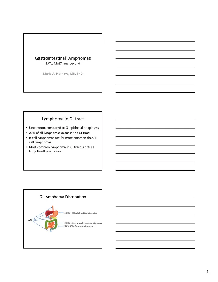
GI Lymphoma Distribution

RARE 55‐65%; 5‐10% of all gastric malignancies 20‐35%; 25% of all small intestinal malignancies 7‐20%; 0.5% of colonic malignancies

slide-2
SLIDE 2

2

Classic Sites of GI Lymphomas

Site Lymphoma Stomach MALT Lymphoma 2nd portion of duodenum Primary intestinal follicular lymphoma Small intestine EATL Terminal ileum Burkitt lymphoma Colonic polyps Mantle cell lymphoma (lymphomatous polyposis)

How to approach lymphoid processes?

  • “SurgPath” / GI view

– What disease could this be:

  • Inflammatory conditions
  • Lymphoma
  • Another malignancy (epithelial, myeloid, mesenchymal)
  • Normal ??

– Immunostains

  • Some “CDs”, other immunos (Keratins, etc.)
  • “Hemepath” view

– Morphology – lymphoid collections are fun! – Immunostains (lots more of “CDs”) – Molecular studies ?? – Conclusions: Lymphoma / Reactive / Atypical

  • Consult the other side at least once

Thoughts to consider

  • Small amount of tissue (usually), but
  • The endoscopist’s description can provide important clues
  • However, the GI tract has normal populations of lymphoid tissues

and can have lots of inflammatory conditions – both can give rise/result in lymphoproliferative disorders and confound our diagnosis of them

https://emedicine.medscape.com/article/175909‐overview SY Min, et al. Clin Endosc. 2013;46(6):647‐650.

slide-3
SLIDE 3

3

Native mucosa‐associated lymphoid tissue (MALT) vs acquired MALT

Peyer’s patch

  • H. pylori gastritis

Tough Decisions

  • Does a “label” of lymphoma lead to appropriate management?
  • Toughest when the process is small and/or early

– Is it really lymphoma?

  • Or inflammatory process?
  • Or normal MALT?

– Endoscopic impression? – How can it be followed? – Should it be treated and how?

Additional complexities:

  • Balance between pragmatic approach and

keen eye for subtle findings

  • Unusual variants and mimics present

conundrums and pitfalls

  • 2016 Update to the WHO Classification of

Tumors of Haematopoietic and Lymphoid Tissue

slide-4
SLIDE 4

4

Some Practical Examples

  • Diffuse Large B‐cell Lymphoma
  • Extranodal Marginal Zone Lymphoma of Mucosa‐

associated Lymphoid Tissue

  • Follicular Lymphoma
  • Mantle Cell Lymphoma
  • Burkitt Lymphoma
  • Enteropathy‐Associated T‐cell Lymphoma
  • Monomorphic Epitheliotropic Intestinal T‐cell

Lymphoma

  • NK/T Lymphoma, nasal type
  • Hepatosplenic T‐cell lymphoma

Diffuse Large B‐cell Lymphoma (DLBCL)

DLBCL

  • Most common type of lymphoma in GI tract
  • Mature large B‐cell lymphoma that can occur

anywhere in GI tract

  • May arise de novo or evolve from underlying low‐

grade B‐cell lymphoma

  • Subtypes related to Epstein‐Barr infection

– Predilection for elderly and immunosuppressed – If arises in iatrogenically immunocompromised following transplant, then classified as monomorphic post‐transplant lymphoproliferative disorder (PTLD)

  • Clinically aggressive

– Potentially curable with chemotherapy and immunotherapy – low‐grade B‐cell component may be refractory and persist

slide-5
SLIDE 5

5

DLBCL: Morphology

10x 10x

400x

DLBCL: Immunophenotype

  • Pan‐B cell marker expression

– CD20, CD79a, Pax‐5

  • Aberrant Bcl‐2 expression (most)
  • Germinal center B‐cell phenotype

– CD10, Bcl‐6 expression

  • Activated B‐cell type

– MUM1/IRF4 expression

  • No expression of T‐cell markers

– except CD5 (~10%)

Hans Algorithm

Hans CP, et al. Blood. 2004;103(1):275‐82

slide-6
SLIDE 6

6

DLBCL: Other Immunophenotypic and Molecular/Cytogenetic Features

  • MYC alterations and expression

– MYC rearranged in 5‐15% of DLBCL, NOS

  • Frequently associated with BCL2 or BCL6 translocation = “double hit”
  • r “triple hit” lymphomas
  • new formal category in WHO2016: High‐grade B‐cell lymphoma

(HGBL) with rearrangements of MYC and BCL2 and/or BCL6

– MYC protein expression in 30‐50% of DLBCL, associated with concomitant BCL2 expression in 20‐35%

  • BUT do not carry MYC/BCL2 chromosomal alteration, thus named

“double expressor lymphoma”

  • Positive expression: at least 40% for c‐myc and 50% for Bcl‐2 by IHC
  • Prognostic indicator: double‐expressor lymphomas have worse
  • utcome than other DLBCL, NOS but are not as aggressive as HGBL

with rearrangements of MYC and BCL2 and/or BCL6

DLBCL: Other Immunophenotypic and Molecular/Cytogenetic Features

  • CD30 expression

– Target for brentuximab vedotin immunotherapy

  • NGS studies

– GCB‐DLBCL: frequent alteration of histone methyl transferase EZH2, BCL2 translocations, and cell motility regulator GNA13 mutations – ABC‐DLBCL: mutations in genes activating BCR/TLR and NFkB pathways (MYD88, CD79a, CARD11, TNFAIP3) – Both: inactivating mutation of TP52, immunosurveillance‐ related genes, alterations in epigenetic regulators, and

  • ncogenic activation of BCL6

Subtype: EBV+ DLBCL, NOS

  • Previously known as EBV+ DLBCL of the elderly
  • In the “elderly” (>50 y): presumed immune

senescence leads to development of lymphoma

– 70% present with extranodal disease (skin, lung, tonsil, stomach) – Aggressive (median survival 2 y)

  • Nicolae et al described a series of EBV+ DLBCL in

young patients (median age 23 y) without known immunodeficiency

– Predominantly nodal disease, 3 of 46 with liver involvement – Good outcome with treatment

slide-7
SLIDE 7

7

Subtype: EBV+ DLBCL, NOS

  • Morphology:

– some resemble T‐cell/Histiocyte‐rich large B‐cell lymphoma with scattered large B cells mimicking HRS cells and variants – some more DLBCL‐like – geographic necrosis common

  • Usually non‐GCB phenotype (CD10‐, MUM1+), EBV+

Nicolae A, et al. Blood. 2015;126(7):863‐72.

CD20 CD79a CD30 PD‐L1 LMP1 EBER IDO (DCs/histiocytes)

Nicolae A, et al. Blood. 2015;126(7):863‐72.

slide-8
SLIDE 8

8

Subtype: EBV+ DLBCL, NOS

  • NOS designation excludes specific EBV‐associated

lymphoma subtypes (Burkitt lymphoma, classical Hodgkin lymphoma, lymphomatoid granulomatosis, primary effusion lymphoma, plasmablastic lymphoma)

  • Implied suggestion to screen cases with above

morphologies for EBV without regard for age

WHO2016 update summary:

Swerdlow SH, et al. Blood. 2016;127(20):2375‐90.

Extranodal marginal zone lymphoma

  • f mucosa‐associated lymphoid tissue

(MALT lymphoma)

slide-9
SLIDE 9

9

MALT lymphoma

  • Mature B‐cell lymphoma that can occur anywhere in GI

tract

– 85% in stomach, often in association with H pylori‐associated gastritis

  • Lymphoma of small mature B lymphocytes that has a

destructive growth pattern (ulcer or thickened mucosal folds)

  • Majority present with low‐stage disease
  • Bone marrow often uninvolved in GI cases
  • M‐proteins are rare, despite relatively frequent

plasmacytic differentiation

– In immunoproliferative small intestinal disease (IPSID), a subtype of MALTL associated with Campylobacter jejuni, a paraprotein is usually found (alpha heavy chain)

MALT Lymphoma: Morphology

  • Reactive germinal centers commonly accompany

lymphoma

– May be invaded or disrupted, leading to “naked” or “moth‐eaten” appearance

  • Heterogeneous, predominantly small B‐lymphocytes

– centrocyte‐like cells (indistinguishable from small cells of normal germinal center – monocytoid cells (slightly larger cells with more ample cytoplasm and slightly indented nuclei) – few scattered large cells (immunoblast‐ and/or centroblast‐like, recapitulate centroblasts of germinal center)

Germinal center Mantle zone Marginal zone

(almost) naked GC moth‐eaten GCs 20x

slide-10
SLIDE 10

10

400x 400x

MALT Lymphoma: Morphology + IHC

  • Some cases have plasma cell differentiation

– Kappa & lambda light chain IHC may be helpful in establishing clonality

  • No distinctive immunophenotype

– Aberrant CD43 expression in only 40‐50% of cases

  • Lymphoepithelial lesion (LEL) is histologic hallmark

– Destructive epithelial infiltration by lymphoma cells – Characteristic but not absolutely specific

CD20 CD3 CD43

slide-11
SLIDE 11

11

Owens SR. Surg Pathol Clin. 2017;10(4):1021‐1037

MALT Lymphoma vs H pylori gastritis MALT Lymphoma: Clinical Aspects

  • 80% are responsive to conservative therapy aimed at

eradication of inciting entity

– Evidence suggests that antibiotic therapy can be effective in H. pylori‐ negative cases of MALT, and in some cases outside the stomach – Gastric MALT lymphomas with t(11;18)(q21;q21) translocation resulting in API2‐MALT1 fusion occur independent of H pylori stimulus and are resistant to conservative therapy

  • Resolution of atypical lymphoid infiltrate can take

months (typically 4‐10 months) to more than a year

– Reporting of residual (regressing) disease on serial biopsies should include comparison statement – Progression is worrisome and requires another treatment modality

MALT Lymphoma: Gray zone cases

  • What to do with borderline or minimal cases??

– “Intense H. pylori gastritis with atypical lymphoid infiltrate” – In the comment address the possibility of early MALTL and offer a statement about typical response to conservative therapy

slide-12
SLIDE 12

12

Follicular Lymphoma Follicular Lymphoma

  • Mature B‐cell lymphoma that may involve the GI

tract secondarily or occasionally primarily

– Duodenal‐type FL presents as multiple small polyps – Formal clinical staging work‐up must be performed

  • CANNOT diagnose primary GI/duodenal‐type FL on histology alone
  • Nodular infiltrate of small mature lymphocytes that

recapitulate follicle center B‐cells (centrocytes and centroblasts)

– Proportions of each population determine grade

  • Typically indolent

– Frequently involve bone marrow and can be difficult to cure – Duodenal‐type FL very indolent, may not need additional therapy beyond local excision

Follicular Lymphoma: Morphology

  • Nodular infiltrate with closely‐packed follicles with

attenuated or absent mantle zones

  • Neoplastic follicles have randomly distributed

centrocytes and centroblasts without tingible‐body macrophages

– In contrast, reactive germinal centers of normal follicles demonstrate polarization due to centrocytes and centroblasts occupation of different zones and have tingible‐body macrophages

slide-13
SLIDE 13

13

20x 100x CD20 BCL‐2

Skinnider BF. Arch Pathol Lab Med. 2018;142(1):44‐52.

CD10

Follicular Lymphoma: Immunophenotype & Cytogenetics

  • Pan‐B cell marker expression (CD20, Pax5)
  • Follicle center cell differentiation (CD10, Bcl‐6)
  • Dendritic cell meshwork present in neoplastic follicles

(highlighted with CD21 and CD23)

  • Aberrant expression of BCL‐2

– Also positive in many other lymphomas and normal T‐cells and plasma cells – Negative in germinal centers of reactive follicles

  • All forms associated with t(14;18)(q32;q21) translocation

involving IGH and BCL2

Mantle Cell Lymphoma

slide-14
SLIDE 14

14

Mantle Cell Lymphoma

  • Systemic small mature B‐cell lymphoma

– Involves GI tract in one‐third of cases – Hepatosplenomegaly, lymphadenopathy, PB involvement common

  • Classically presents as lymphomatous polyposis

– Multiple (sometimes hundreds) of polyps throughout GI tract

  • Aggressive, with overall survival of 3‐5 years

Mantle Cell Lymphoma: Morphology

  • Monomorphic lymphoid proliferation

– Pattern can be diffuse, nodular, “mantle zone”

  • Mantle zone pattern has central follicle surrounded by neoplastic

cells

  • small to medium‐sized neoplastic lymphoid cells with

dark angulated nuclei

  • interspersed hyalinized small vessels (thick‐walled

capillaries) and epithelioid eosinophilic histiocytes (mimicking “starry sky” appearance)

  • Blastoid and pleomorphic variants may mimic ALL

and DLBCL

– Important to recognize as the latter two are potentially curable, whereas MCL is not

400x

slide-15
SLIDE 15

15

Pleomorphic MCL

400x

Classical Mantle Cell Lymphoma: Immunophenotype

  • Pan‐B cell marker expression (CD20, Pax5)
  • Aberrant expression of CD5 (rarely negative), CD43

(usually), BCL‐2, nuclear CyclinD‐1 (very rare negative cases express cyclin D2 or cyclin D3)

  • Surface IgM and/or IgD expression
  • Sox11 expression
  • Negative for CD10, BCL‐6, CD23

Classical Mantle Cell Lymphoma: Cytogenetics

  • Ig genes

– IgH rearranged – IgH Variable regions unmutated or minimally mutated

  • t(11;14)(q13;q32) rearrangement

– Involves IgH and CyclinD1 genes (PRAD1, BCL1) – Classical cytogenetics detects 70‐95% – FISH detects ~100%

  • Other

– p53, p16, p18 (especially in blastoid variant) – 13q14 deletion – Total or partial trisomy 12

slide-16
SLIDE 16

16

MCL: WHO2016 Update

Swerdlow SH, et al. Blood. 2016;127(20):2375‐90.

Burkitt Lymphoma Burkitt Lymphoma

  • Mature B‐cell lymphoma of children and young adults
  • Has very short doubling time
  • Presents often in extranodal sites or as acute leukemia

– Classic lesion is large and destructive mass in distal ileum and/or cecum – Can involve any portion of GI tract

  • Variable global distribution

– Endemic: equatorial Africa and Papua New Guinea – Sporadic: around globe

  • Epstein‐Bar virus association

– Endemic: majority of neoplastic cells in all patients – Sporadic: 30% of cases – Immunodeficiency‐associated (HIV): 25‐40% of cases

  • Good prognosis (up to 90% survival) with appropriate

therapy

slide-17
SLIDE 17

17

Burkitt Lymphoma: Morphology

  • Low magnification: “starry sky”

– Sheets of lymphoma cells are punctuated by tingible‐body macrophages with cellular debris

  • Lymphoma cells are monotonous, medium‐sized,

with round nuclei, dispersed chromatin, inconspicuous nucleoli, scant basophilic cytoplasm

  • Nearly 100% proliferative fraction, numerous mitotic

figures, lots of apoptotic debris

400x

Burkitt Lymphoma: Immunoprofile & Cytogenetics

  • B‐cell marker expression (CD20, CD19, CD79a)
  • Germinal center cell differentiation (CD10, Bcl‐6)
  • High Ki‐67 proliferative index (nearly 100%)
  • Negative for Bcl‐2 (weakly positive in 20%), TdT, CD5
  • MYC translocation is characteristic (but not specific)

– t(8;14)(q24;q32): c‐MYC and IgH (75%) – t(2;8)(p12;q24): Ig kappa and c‐MYC (15%) – t(8;22)(q24;q11): c‐MYC and Ig lambda (10%)

slide-18
SLIDE 18

18

Enteropathy‐Associated T‐cell Lymphoma (EATL)

EATL

  • Aggressive T‐cell lymphoma that produces large,

destructive masses, often in jejunum

  • Arises in patients with celiac disease

– In setting of refractory sprue – As sentinel event in patients with undiagnosed celiac disease

  • Associated with HLA haplotypes DQ2 and DQ8

– Northern European descent

  • Poor prognosis due to aggressive nature and

debilitated state of patients with malabsorption

– Common presentation is ulcerated mass +/‐ perforation – Median survival of months

EATL: Morphology

  • Diffuse, destructive infiltrate of intermediate‐sized or

large cells with angulated or pleomorphic nuclei (resembling those of DLBCL) with prominent nucleoli

  • Tumor infiltration by inflammatory cells including

histiocytes and eosinophils

  • Neoplastic cells infiltrate individual crypts
  • Areas of necrosis may be present
  • Adjacent intestinal mucosa demonstrates variable

degree of enteropathy (villous atrophy, crypt hyperplasia, intraepithelial lymphocytosis)

slide-19
SLIDE 19

19

5x 60x 400x

Arps DP, Smith LB. Arch Pathol Lab Med. 2013;137(9):1227‐31. Delabie J, et al. Blood. 2011;118(1):148‐55.

250x

EATL: Immunophenotype

  • Cytotoxic phenotype (granzyme B, TIA‐1)
  • TCR alpha/beta in most cases
  • Typically positive for CD3, CD7, occasional cases

CD8+, variable CD30

  • Usually negative for CD4, CD8, CD5, CD56, MATK
  • Intraepithelial lymphocytes in the adjacent

enteropathic mucosa have a similar phenotype

Monomorphic Epitheliotropic Intestinal T‐cell Lymphoma (MEITL)

Formerly known as EATL II or EATL, monomorphic form

slide-20
SLIDE 20

20

MEITL

  • Aggressive T‐cell lymphoma
  • NO association with celiac disease
  • Apparent increased frequency in patients of Asian

and Hispanic descent

  • Poor prognosis due to aggressive nature and

debilitated state of patients

– Common presentation is ulcerated mass +/‐ perforation – Median survival of months

MEITL: Morphology

  • Diffuse, destructive, often ulcerated infiltrate of

monotonous medium‐sized lymphoid cells with dispersed chromatin, inconspicuous nucleoli, and ample clear cytoplasm (monocytoid appearance)

  • Prominent epitheliotropism of tumor cells in

adjacent mucosa with little involvement of submucosa/muscularis propria (lateral spread)

  • Distant mucosa without enteropathy
  • Paucity of reactive inflammatory cells within tumor
  • Tumor perforation frequent

Tan SY, et al. Leukemia. 2013;27(8):1688‐96.

slide-21
SLIDE 21

21

MEITL: Immunophenotype

  • Cytotoxic phenotype (TIA‐1, granzyme B)
  • Typically express CD2, CD3, CD7, CD8, CD56, nuclear

MATK

  • TCR gamma/delta in most cases

– Some TCR silent – Some TCR alpha/beta

  • Typically negative for CD5, CD4

EATL MEITL Frequency 80‐90% 10‐20% Epidemiology Complication of GSE Occurs sporadically associated with HLA‐DQ2/DQ8 refractory GSE patients at high risk Northern Europeans descent Asian and Hispanic descent Morphology Variable, pleomorphic, intermediate to large cells Monotonous small to intermediate‐sized cells Angulated nuclei Round nuclei Prominent nucleoli Inconspicuous nucleoli Areas of necrosis Rare necrosis Variable to heavy background mixed inflammatory infiltrate Minimal background inflammatory infiltrate Immunophenotype CD3+, CD5‐, CD7+ CD3+, CD5‐, CD7+ CD8‐ (80%) CD8+ (80%) CD56‐ (>90%) CD56+ (>90%) nuclear MATK‐ nuclear MATK+ Cytogenetics +9q31.3 or ‐16q12.1 86% 83% +1q32.2‐q41 73% 27% +5q34‐q35.2 80% 20% +8q24 (MYC) 27% 73%

Extranodal Natural Killer/T‐cell Lymphoma, Nasal Type (ENKTL)

slide-22
SLIDE 22

22

ENKTL

  • Rare, very aggressive lymphoma most commonly

involving upper aerodigestive tract

– Propensity to involve the GI tract

  • Frequent tumor ulceration due to angiocentric and

angiodestructive growth

  • Associated with Epstein‐Barr virus
  • Higher prevalence in Asian and Native American

populations

  • Variable prognosis for nasal ENKTL, but extranasal ENKTL

has short survival times and poor response to therapy

ENKTL: Morphology

  • Variably‐sized neoplastic cells

– Small, medium‐sized, large, or anaplastic – Irregularly folded nuclei with granular or vesicular chromatin – Inconspicuous nucleoli – Moderate pale cytoplasm

  • Mitotic figures easily seen
  • Angiocentric and angiodestructive growth with

fibrinoid changes in vessels

  • Coagulative necrosis and many apoptotic bodies

Owens SR. Surg Pathol Clin. 2017;10(4):1021‐1037

slide-23
SLIDE 23

23

ENKTL: Immunophenotype

  • Typically express CD2, CD56, CD3epsilon

(cytoplasmic), cytotoxic molecules (granzyme B, TIA1, perforin), CD43, CD25

  • EBV+ (by IHC or ISH)
  • Typically negative for surface CD3, CD4, CD8, CD5,

CD16, CD57

  • TCR in germline configuration

Hepatosplenic T‐cell Lymphoma (HSTL)

HSTL

  • Rare extranodal, systemic cytotoxic gamma/delta T‐

cell lymphoma of young adults

  • 20% arise in setting of chronic immune suppression

– After solid‐organ transplant; considered PTLD – Immunosuppression for IBD

  • Presents with marked splenomegaly, (usually)

hepatomegaly, without lymphadenopathy, but with BM involvement

– Marked thrombocytopenia – Often anemia, leukopenia

  • Aggressive, with relapses after treatment is most cases

– Median survival <2 years

slide-24
SLIDE 24

24

HSTL: Morphology

  • Spleen: diffuse involvement of cords and sinuses of

red pulp, white pulp atrophy

  • Liver: diffuse infiltration of sinusoids
  • Neoplastic cells are monotonous medium‐sized cells

with pale cytoplasm

100x 400x

slide-25
SLIDE 25

25

400x

HSTL: Immunophenotype

  • Typically express CD3, CD2, CD56 (frequent), TCR

gamma/delta (alpha/beta in a minority), and cytotoxic granule‐associated proteins (TIA1 and granzyme M)

  • Typically negative for CD4, CD8 (minority +), CD5 &

CD7 (frequent loss), granzyme B, perforin, CD57, CD30

Approach to Evaluation

  • Careful morphologic evaluation

– Inflammatory conditions vs Lymphoma vs Another malignancy vs Normal

  • Tiered immunostain panels

– CD20, CD3, CD43

  • Reactive: T‐cell predominant, but mixed
  • Aberrant CD43 expression on CD20‐positive B‐cells: MCL, CLL/SLL,

subset of MALTL

  • CD43 expression without CD20 or CD3: possibility of myeloid neoplasm

– Targeted additional immunos as needed to complete characterization of an entity or exclude others – Targeted additional immunos as needed for prognosis or treatment

  • Molecular studies for specific gene alterations if needed

for diagnostic refinement

  • Clonality studies may not be useful

– Benign reactive populations can have small clones that may amplify erroneously leading to lymphoma diagnosis

slide-26
SLIDE 26

26

Quick Case Study

  • Elderly Caucasian man
  • Celiac sprue x 7 years
  • Presented with abdominal pain, nausea, and

vomiting

  • Imaging revealed partially obstructing duodenal

mass

2x flattened villi ulcer

Duodenal biopsy:

4x

slide-27
SLIDE 27

27

Low Power DDx

  • Gluten‐sensitive enteropathy / celiac disease

– Refractory GSE – Collagenous sprue

  • Tropical sprue
  • Totally flat mucosa rare in tropical sprue
  • Autoimmune enteropathy
  • Medication effect

– NSAIDs – Olmesartan – Colchicine – Mycophenolate mofetil – Ipilimumab (anti‐CTLA4) – Chemotherapy agents

  • Inflammatory bowel disease

200x 600x

slide-28
SLIDE 28

28

400x

Immunophenotype

CD3 CD56 CD8 CD4 CD5 CD7

EATL MEITL Frequency 80‐90% 10‐20% Morphology Variable Monomorphic small to medium Immunophenotype CD8 Mostly negative (20%+) Mostly positive (80%+) CD56 Negative (>90%) Positive (>90%) HLA‐OQ2/‐0Q8 Positive (>90%) Positive (30‐40%) antecedent GSE present absent GSE changes in adjacent mucosa Villous atrophy, crypt hyperplasia, lamina propria lymph‐ & plasmacytosis, IELs Villous atrophy, crypt hyperplasia, lamina propria w/o inflammatory background, IELs

Diagnosis: EATL

slide-29
SLIDE 29

29

References:

  • Nicolae A, et al. EBV‐positive large B‐cell lymphomas in young patients: a nodal lymphoma with evidence for a

tolerogenic immune environment. Blood. 2015;126(7):863‐72.

  • Asano N, et al. Helicobacter pylori‐negative gastric mucosa‐associated lymphoid tissue lymphomas: A review.

World J Gastroenterol. 2015;21(26):8014‐20.

  • Nomura E, et al. Regression of MALT lymphoma of the rectum after Helicobacter pylori eradication therapy in a

patient negative for Helicobacter pylori. Nihon Shokakibyo Gakkai Zasshi. 2010;107(9):1466‐73.

  • Nakamura S, et al. Helicobacter pylori‐negative gastric mucosa‐associated lymphoid tissue lymphoma: a

clinicopathologic and molecular study with reference to antibiotic treatment. Cancer. 2006;107(12):2770‐8.

  • Owens SR. Lymphoproliferative Diseases of the Gut: A Survival Guide for the General Pathologist. Surg Pathol
  • Clin. 2017;10(4):1021‐1037.
  • Swerdlow SH, et al. The 2016 revision of the World Health Organization classification of lymphoid neoplasms.
  • Blood. 2016;127(20):2375‐90.
  • Skinnider BF. Lymphoproliferative Disorders of the Gastrointestinal Tract. Arch Pathol Lab Med. 2018;142(1):44‐52.
  • Arps DP, Smith LB. Classic versus type II enteropathy‐associated T‐cell lymphoma: diagnostic considerations. Arch

Pathol Lab Med. 2013;137(9):1227‐31.

  • Delabie J, et al. Enteropathy‐associated T‐cell lymphoma: clinical and histological findings from the international

peripheral T‐cell lymphoma project. Blood. 2011;118(1):148‐55.

  • Tan SY, et al. Type II EATL (epitheliotropic intestinal T‐cell lymphoma): a neoplasm of intra‐epithelial T‐cells with

predominant CD8αα phenotype. Leukemia. 2013;27(8):1688‐96.

  • Shi Y, Wang E. Hepatosplenic T‐Cell Lymphoma: A Clinicopathologic Review With an Emphasis on Diagnostic

Differentiation From Other T‐Cell/Natural Killer‐Cell Neoplasms. Arch Pathol Lab Med. 2015;139(9):1173‐80. Review.

  • J Dig Dis. 2015;16(4):169‐76.
  • Peng JC, Zhong L, Ran ZH. Primary lymphomas in the gastrointestinal tract. J Dig Dis. 2015;16(4):169‐76.

Questions?