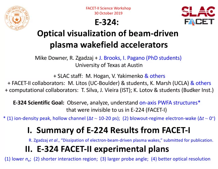

FACET-II Science Workshop 30 October 2019 E-324: Optical visualization of beam-driven plasma wakefield accelerators Mike Downer, R. Zgadzaj + J. Brooks, I. Pagano (PhD students) University of Texas at Austin + SLAC staff: M. Hogan, V. Yakimenko & others + FACET-II collaborators: M. Litos (UC-Boulder) & students, K. Marsh (UCLA) & others + computational collaborators: T. Silva, J. Vieira (IST); K. Lotov & students (Budker Inst.) E-324 Scientific Goal: Observe, analyze, understand on-axis PWFA structures* that were invisible to us in E-224 (FACET-I) * (1) ion-density peak, hollow channel (∆ t ~ 10-20 ps); (2) blowout-regime electron-wake (∆ t ~ 0 + ) I. Summary of E-224 Results from FACET-I R. Zgadzaj et al ., “Dissipation of electron-beam-driven plasma wakes,” submitted for publication. II. E-324 FACET-II experimental plans (1) lower n e ; (2) shorter interaction region; (3) larger probe angle; (4) better optical resolution
In FACET-I, we imaged near-field diffraction patterns of an ion wake in a single shot heat-pipe oven θ = 8 mrad e- ptychography? 1.5 m bunch E e = 20 GeV - z 1 0 + z 1 Q = 2.4 nC σ r = 30 µm σ z = 55 µm Li vapor f /40 lens n a = 0.8e17 cm -3 E pr = 1 mJ λ pr = 0.8 µm CCD ∆t σ r = 0.5 cm probe τ pr = 0.1 ps image plane jitter ~ 0.1 ps 2 normalized intensity [arb. units] 2 0.03 ns 0.3 ns ∆t < 0 radial position 0 [mm] 2 1 2 10 µs 0.9 ns 0.6 ns 0 2 0 0 0 -30 30 -30 30 -30 0 30 longitudinal position in heat-pipe oven [cm]
LCODE-simulated n e (r) profiles reconstruct observed fringe patterns at ∆t ≥ 0.1 ns MEASURED CALCULATED calculations by K. V. Lotov, including impact ionization original ions only V. K. Khudyakov, A. Sosedkin 2 2 equivalent radius in interaction region [mm] 0 100 ps [arb. units] 2 2 0 300 ps 2 1 2 0 normalized intensity 600 ps 2 2 0 900 ps 0 2
Our FACET-II scientific goal: observe, analyze & understand on-axis PWFA structures* FACET-I geometry planned for FACET-II * 1) At ∆t ~ 10-20 ps: on-axis ion density peak, quasi-hollow channel favorable for focused positron accleration • A. A. Sahai, Phys. Rev. Accel. Beams 20 , 081004 (2017) • T. Silva, J. Vieira et al ., forthcoming presentation at this workshop. * 2) At ∆t < 1 ps: strongly nonlinear electron wake b) e - vs. e + driven wakes a) pre-ionized vs. self-ionized plasma c) transverse wake instabilities: dependence on drive bunch shape, pre-formed plasma
FACET-II’s shorter Li oven & lower n e enable steeper θ probe , optical access to interior plasma structures 2.34 m Bellows angle when bypass pipe is used during alignment 6” cube FACET I Bypass pipe 3” beam-ionized Li ( n e = 8e17 cm -3 ) Oven pipe Cooling Gate f/# 60 jacket valve Pipe wall (3.175cm) 3.81cm 1.125” 1.25” limiting optical aperture 1.22 m wick Oven pipe transverse dimensions the same for FACET I and II 8” cube Bypass pipe 4” Bellows angle when oven pipe Oven pipe is used during experiments FACET II f/# 25 laser pre-ionized Li ( n e = 5e17 cm -3 , r p ~ 200 µm) Enlarged output cube (6” à 8”) & output mirror (3” à 4” diam.) improve probe collection efficiency. Lower f # lens (60 à 25) improves imaging resolution.
Simulated probe images show signatures of evolving on-axis ion density maxima • laser pre-ionized Li plasma • r p ≈ 200 µm • λ pr = 800 nm • n e = 3.5 x 10 16 cm -3 t 3 ≈ 22 ps t 2 ≈ 16 ps t 1 ≈ 7 ps • θ pr = 20 mrad 200 OSIRIS plasma simulations by T. Silva, IST on- axis peak light 0 interior hollow channel 200 µm probe simulations by R. Zgadzaj, UT-Austin Li refractive index
Probe at θ pr = 8 mrad less sensitive to on-axis plasma structures • laser pre-ionized Li plasma • r p ≈ 200 µm • λ pr = 800 nm • n e = 3.5 x 10 16 cm -3 t 1 ≈ 7 ps t 2 ≈ 16 ps t 3 ≈ 22 ps • θ pr = 8 mrad 200 OSIRIS plasma simulations by T. Silva, IST dark 0 interior 200 µm probe simulations by R. Zgadzaj, UT-Austin Li refractive index
Shape of plasma column edge influences how on-axis structures modify probe image • laser pre-ionized Li plasma • r p ≈ 200 µm • λ pr = 800 nm • n e = 3.5 x 10 16 cm -3 t 1 ≈ 7 ps • θ pr = 20 mrad 200 OSIRIS plasma simulations by T. Silva, IST 0 Image of probe (∆ t ≈ +20 ps) can be normalized to 200 200 µm image of reference pulse (∆ t ≈ -1 ps) to correct for this. probe simulations by R. Zgadzaj, UT-Austin plasma edge: super- plasma edge: super- Gaussian order 20 Gaussian order 8 Li refractive index
• laser pre-ionized Li plasma • r p ≈ 200 µm • λ pr = 800 nm • n e = 3.5 x 10 16 cm -3 t 1 ≈ 7 ps • θ pr = 20 mrad 200 OSIRIS plasma simulations by T. Silva, IST 0 200 µm simulated reference pulse image probe simulations by R. Zgadzaj, UT-Austin plasma edge: super- Gaussian order 8 Li refractive index
At 0.5 > ∆t > 0.2 ps, probe remains within 1 st bucket of electron wake for 20 cm Geometric walkoff @ θ pr = 20 mrad: +40 µm probe Group-velocity walkoff: -2 µm Walk-off ~38μm 20 mrad e-bunch e-bunch Overlap region ~ 20cm* probe * for 4 mm wide probe (4σ diameter) and wake axis @ θ pr = 20 mrad
Under FACET-II conditions, a fully-blown-out e-beam-driven wake will be visible • laser pre-ionized Li plasma • r p ≈ 200 µm • λ pr = 800 nm bkgd. plasma bkgd. plasma • n e = 3.5 x 10 16 cm -3 only; no wake • θ pr = 20 mrad + electron wake ∆ t ≈ 0.3 ps 200 fully-blown-out bubble 0 200 µm probe simulations by R. Zgadzaj, UT-Austin Li refractive index
The simulations motivate additional experimental upgrades for PWFA imaging PROBE independent of ionizing laser UPGRADES: PROBE (1) double θ pr à probe interior plasma structures (2) reduce f # à higher image resolution θ pr = a (3) install NL phase con- L p = Plasma oven length 20 mrad trast optics à improve sensitivity to n e < 10 16 cm -3 (4) install multiple image- object planes + reference à distinguish on-axis from edge structures. ionizing laser beam apre-tested in our U. Texas lab FS CCD 3 CCD 2 CCD 1 L1 Li, Opt. Lett. 38 , 5157 (2013) phase- contrast multiple image planes
Later FACET-II Experiments He/H 2 chamber with transverse optical access will enable Faraday profiling* of magnetized PWFAs * LWFA expts.: Kaluza, PRL 105 , 11502 (2010); Buck, Nat. Phys . 7 , 453 (2011) [ n e > 10 19 cm -3 ] ~ longitudinal Chang, PhD (2018) [ n e ~ 10 17 cm -3 ] probe UPGRADES: transverse probe (1) double θ pr à probe interior plasma structures (2) reduce f # à higher image resolution (3) install NL phase con- trast optics à improve sensitivity to n e < 10 16 cm -3 CCD Cotton- (4) install multiple image- Faraday ro- Mouton object planes + reference tation optics optics à distinguish on-axis from CCD ! ! ! ! k pr ⊥ edge structures. B φ k pr || B φ b (5) install Faraday/C-M probes à sensitive profil- probe of: (1) bubble sheath structure & dynamics ing of low- n e structures bpre-tested in Texas PW expts. (2) external or Trojan horse e - -injection Chang, PhD dissertation (2018)
Faraday rotation picks out dense bubble wall in tenuous plasma Based on technique developed by: Kaluza, PRL 105 , 115002 (2010) ; Buck, Nat. Phys . 7 , 453 (2011) in n e > 10 19 cm -3 plasma ; results shown from Chang, PhD dissertation, UT-Austin (2018) Faraday probe setup Faraday rotation results Texas PW probe pump probe 2 GeV 100 J, 150 fs pump e - e-bunch 10 cm gas cell ~10cm Gas cell ( n e0 = 2•10 17 cm -3 ) anamorphic Anamorphic Imaging System imaging CCD CCD polarizer Cubic Polarizer polarizer Cubic Polarizer CCD CCD • separation of ± lobes: bubble size 4 • |∆ ϕ Faraday |: bubble wall density measure- • width of each lobe: bubble wall thickness ments • longitudinal variations: bubble evolution
SUMMARY • In FACET-II, we will visualize on-axis e- and ion-wake structures, taking advantage of larger θ pr , higher-resolution imaging, lower n e than in FACET-I. ∆ t ≥ 30 ps ∆ t < 1 ps (1) on-axis ion-density peak (3) e-wake structure & propagation dynamics (2) hollow-channels (4) transverse & longitudinal e-wake instabilities • Our simulations show that our probe will illuminate these structures well, and provide clear optical signatures of them... • ... but there are challenges. Probe images are... ... low contrast à use phase-contrast imaging ... convolved with plasma-edge signatures à use probe-reference pulse pair.
II. Overview of FACET-I results (E-224) OSIRIS 1 simulations show nonlinear e-wake excitation & early (∆t < 40 ps) ion response e-bunch density [e cm -3 ] simulations by T. Silva, J. Vieira (IST) 10 15 10 16 10 17 10 18 plasma density n e [10 17 cm -3 ] OSIRIS 1 framework 2 incident e-bunch Massivelly Parallel, Fully · Relativistic PIC Code Visualization & Data · 1 Analysis Infrastructure moving window ionization & NL e-wake excitation Developed by Osiris Con- · sortium, UCLA + IST 0 1 Fonseca et al ., PPCF 55 , 124011 (2013) fixed window ∆ t = 40ps E r ( z ) (∆ t = 1 ps) < E r > 2 experiment radial distance [mm] 0 2 fixed window reconstruction 2 ρ Li+ ( z ) (∆ t = 40 ps) < ρ Li+ > 0 2 -20 0 20 longitudinal distance [cm]
Recommend
More recommend