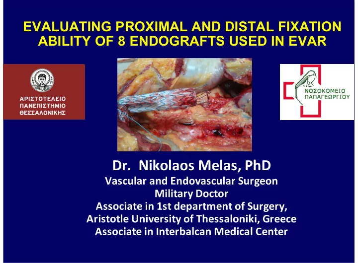

EVALUATING PROXIMAL AND DISTAL FIXATION ABILITY OF 8 ENDOGRAFTS USED IN EVAR Dr.$$Nikolaos$Melas,$PhD Vascular$and$Endovascular$Surgeon Military$Doctor Associate$in$1st$department$of$Surgery, Aristotle$University$of$Thessaloniki,$Greece Associate$in$Interbalcan$Medical$Center
From the very beginning of EVAR introduction, with tube endografts, Parodi 1990 (1) 1. Parodi JC, Palmaz JC, Barone HD. Transfemoral intraluminal graft implantation for AAA. Ann Vasc Surg 1991; 5 :491-9 2. Parodi JC, Barone A, Piraino R, Schonholz. Endovascular treatment of abdominal aortic aneurysms: lessons learned. J Endovasc Surg 1997;4: 102-10
Lifepath Vanguar Ancur Corvit Cordis AneuR Exclude Zenith PowerLin Talen Fortran Appol d e a x r k t o Aorfix Anaconda Enduran Aptus t Or later bifurcated systems
It became clear that for durable sac exclusion, there should exist adequate Fixation zones Sealing zones In order to achieve uncomplicated long-term results
Established Fixation Methods Endografts Suspension, SR, Hooks, Radial Force Columnar strength Bards, Anchors Ø Are not sewn Ø Are not incorporated Ø require continuous mechanical fixation in order to withstand pulsatile blood forces (1,2) This is achieved by radial force : • Blood pressure, oversizing producing friction columnar strength • suspension (SR stent, barbs, hooks, • anchors, pins, proximal stent frixion) Malina M, et al. Endovascular healing is inadequate for fixation of Dacron stent grafts in human aorta ilial vessels. Eur J Vasc Endovasc Surg. 2000; 19: 5–11. Zarins CK. Stent-Graft Migration: How Do We Know When We Have It and What Is Its Significance. JEVT 2004;11:364–365.
Loss of fixation - consequences Sac re- Migration Endoleak I pressurization Rupture Greenberg RK, et al. Stentgraft migration: a reappraisal of analysis methods and proposed revised definition. J Endovasc Ther. 2004;11:353–363. 1. Luis R. Leon, Jr and Heron E. Rodriguez. Aortic Endograft Migration. Perspectives in Vascular Surgery and Endovascular Therapy. 2005, Volume 17, Number 4, 363-373. Conners MS, Sternbergh WC, Carter GS, Tonnessen BH, Yoselevitz M, Money SR, et al. Secondary procedures following endovascular aneurysm repair. J Vasc Surg. 2002;36:992–996. Ivancev K, Malina M, Lindbland B, et al. Abdominal aortic aneurysms: Experience with the Ivancev-Malmo endovascular system for for aortomonoiliac stent graft. J Endov Surg. 1997; 4 :242-251. Malina M, et al. Endovascular healing is inadequate for fixation of Dacron stent grafts in human aorta ilial vessels. Eur J Vasc Endovasc Surg. 2000; 19: 5–11. Zarins CK. Stent-Graft Migration: How Do We Know When We Have It and What Is Its Significance. JEVT 2004;11:364–365. Liffman Κ , Et al. Analytical Modeling and Numerical Simulation of Forces in an Endoluminal Graft. JEVT. 2001;8:358–371. 9. White G, et al. “Endoleak” a proposed new terminology to describe incomplete aneurysm exclusion by an endoluminal graft. J Endovasc Surg1996; 3 : 124–125. 10. Veith FJ, et al. Nature and significance of endoleaks and endotension: summary of opinions expressed at an international conference. J Vasc Surg 2002;35:1029-35. 11. Conners MS 3rd, et al. Endograft migration one to four years after endovascular abdominal aortic aneurysm repair with the AneuRx device: a cautionary note. J Vasc Surg, 2002; 36:476-484. 12. Luis R. Leon, et al. Aortic Endograft Migration. Perspectives in Vascular Surgery and Endovascular Therapy. 2005, Volume 17, Number 4, 363-373.
Migration - definition • Endograft movement >10 mm in relation to fixed anatomic landmarks as SMA or renals (for proximal) and IIA for distal. (1) Rare Immediate (2-4) Perioperative or Within 30 days Due to wrong indication for suitable anatomy /graft choice, or technical insufficiency Late (2-4) More often After 30 days, usually after the 1st year increasing frequency thereafter Due to neck dilatation / remodeling, endoleak I, material fatigue Main pathophysiology •The continuous force applied by the pulsatile blood flow against the graft which is not incorporated to the aortic wall but needs permanent mechanical fixation (anchoring, suspension, radial force) to remain stable. (5,6) Greenberg RK, et al. Stentgraft migration: a reappraisal of analysis methods and proposed revised definition. J Endovasc Ther. 2004;11:353–363. 1. Luis R. Leon, Jr and Heron E. Rodriguez. Aortic Endograft Migration. Perspectives in Vascular Surgery and Endovascular Therapy. 2005, Volume 17, Number 4, 363-373. Conners MS, Sternbergh WC, Carter GS, Tonnessen BH, Yoselevitz M, Money SR, et al. Secondary procedures following endovascular aneurysm repair. J Vasc Surg. 2002;36:992–996. Ivancev K, Malina M, Lindbland B, et al. Abdominal aortic aneurysms: Experience with the Ivancev-Malmo endovascular system for for aortomonoiliac stent graft. J Endov Surg. 1997; 4 :242-251. Malina M, et al. Endovascular healing is inadequate for fixation of Dacron stent grafts in human aorta ilial vessels. Eur J Vasc Endovasc Surg. 2000; 19: 5–11. Zarins CK. Stent-Graft Migration: How Do We Know When We Have It and What Is Its Significance. JEVT 2004;11:364–365.
Purpose •evaluate the differences of proximal, distal and overall fixation mechanisms within 8 commercially available endografts Validate various parameters that might influence fixation. Melas N, Saratzis A, Saratzis N, Lazaridis J, Psaroulis D, Trygonis K, Kiskinis D. Aortic and iliac fixation of endografts for abdominal-aortic aneurysm repair in an experimental model using human cadaveric aortas. Eur J Vasc Endovasc Surg. 2010 Oct;40(4):429-35.
Methods •20 human cadaveric aortas •Mean proximal infrarenal aortic diameter 20,5 mm (range 19,2-21,9) PTFE prosthesis as control Endofit Talen Zenit Enduran VI BE t h t Cuff Anaconda Excluder Powerlink Validated Endografts
Methods Cadaveric preperation
Abdominal aorta was exposed, and pressurization followed for OD measurement
Methods Aortas were surgically dissected from renals to iliac bifurcations, left in situ and transected 2 cm below the renals and above aortic bifurcation, to mimic AAAs’ proximal and distal landing zones SMA RENALS NECK IMA IVC ILIACS Aortoiliac dissection
Methods CL limb Main body Endografts were inserted
Methods 1 2 3 4 Deployed in the usual manner
Methods •Grafts were connected via a strong suture (kevlar cord) to a force gauge •Caudal force was applied to the flow divider of each graft. 5 6 Kevlar cord
Methods Recordings were repeated without iliac fixation Similar protocol was applied for iliac limbs but the DF was cephalad Recordings were repeated after molding balloon dilatation
Methods The pull out force recorded until dislocation from fixation zone was defined as displacement force (DF) DF DF
Powerlink Endurant Excluder Zenith
Endofit AUI Anaconda Vascular Innovation Talent Veith BE Cuff
Results Statistics •Shapiro Wilk test ,Kolmogorov Smirnov test. •Mann-Whitney U test (non parametric data) •Student’s T-test (parametric data). •p<0,05 significant .
We(acuired((8((different(categories Results TALENT ANACΟNDA EXCLUDER ENDOFIT(AUI ZENITH ENDURANT POWERLIN HAND K SEWN 1)(Endograft(fully(deployed 14.90 28.75 17.90 12.15 34,50 26.75 13.65 &&& Validated Overall fixation (14.40& (26.50,&,31.05) (17.30&18.85) (11.00&13.40) (31.35&37.50) (24.60& (12.50&14.90 without(molding(balloon 15.30) 28.70) ) against caudal migration 2)(Endograft(fully(deployed 16.20 36,10 22.60 13.10 39.20 31.70 14,80 76.20 (15.70& (34.90&37.50) (21.85&23.30) (12.50&14.00) (37.80&40.90) (29.50& (14.10& (66.40 after(molding(balloon 16.65) 34.05) 15.50) 79.00) 3)(Only(body(deployed 8.20 27.95 14.30 12.10 32.05 25.50 6.50 &&& (7.05&9.25) (25.05&30.85) (13.40&15.40) (11.70&12.40) (29.65&34.60) (23.95& (6.45&6.70) without(molding(balloon 27.05) Validated Proximal fixation against caudal migration 4)(Only(body(deployed 9.10 35,70 18.00 12.25 36.80 30.10 7.10 &&& (8.30&9.95) (34.65&36.80) (16.80&18.30) (11.20&13.05) (34,70&38.75) (26.30& (7.00&7.25) after(molding(balloon 34.20) 5)(Iliac(leg(deployed(2(cm 6.85 8,90 7,65 6.75 7.15 7,30 2,65 &&& (6.40&7.30) (7.75&9.90) (7.20&8.10) (5.00&7.10) (6.80&7.50) (7.10&7.55) (2.60&3.50) into(iliac(artery without(molding(balloon 6)(Iliac(leg(deployed(2(cm 7.30 9,85 8.05 7.10 7.75 7.85 2,80 &&& (7.00&7.55) (9.55&10.20) (7.30&8.75) (6.00&7.15) (7.25&8.20) (7.15&8.50) (2.70&3.60) into(iliac(artery after(molding(balloon Validated Distal fixation against cephalad migration 7)(Iliac(leg(deployed(5(cm 8.65 13.05 9.90 8.90 9.05 9,05 4,50 &&& (7.55&9.80) (12.15&14.10) (9.45&10.40) (8.05&9.10) (7.55&10.60) (8.50&9.80) (4.35&4.95) into(iliac(artery without(molding(balloon 8)(Iliac(leg(deployed(5(cm 9.20 14.50 10.55 9.00 9.50 9.60 4.75 60.40 (8.00&10.50) (13.95&15.30) (10.10&10.90) (8.30&9.20) (8.05&11.10) (9.25&10.10) (4.55&5.50) (53.50 into(iliac(artery 62.70) after(molding(balloon All(values(refer(to( DF( (displacement(force)(in(( Newton( after(statistical(analysis.(( Median (Z( range ).
Recommend
More recommend