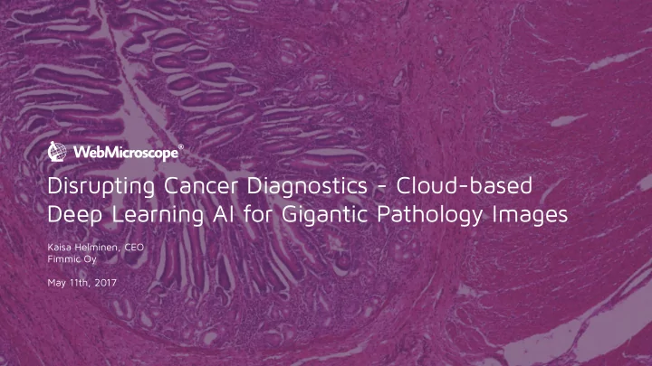

Disrupting Cancer Diagnostics - Cloud-based Deep Learning AI for Gigantic Pathology Images Kaisa Helminen, CEO Fimmic Oy May 11th, 2017
Cancer Every third person affected 14 Million new patients in 2012 +50% more by 2030 Increasing number of samples
Problem Increasing number of samples Gap! Lack of pathologists
Microscopy in cancer diagnostics Subjective, manual analysis Risk for misdiagnosis and wrong medication Agreement can be as low as 48%
Digitalization -> Deep Learning AI
Challenges Gigapixel-sized files (Gb-Tb) Locked image file formats Image Analysis software are expensive on-premise installations Conventional machine vision technology is limited designed by Freepik
Artificial WebMicroscope Intelligence Workflow Any microscope Pathologists Researchers Educators Samples Any device scanner
Artificial Intelligence & Deep Learning Facial recognition Self-driving cars Tissue diagnostics id-labs.org The Guardian
Different types of Deep Learning Image Analysis tasks 1. Laborious quantification tasks, combined with ROI selection, e.g. quantification of certain cells 2. Segmentation of tissue based on morphology, e.g. tumor grading, epithelium/stroma segmentation 3. Detecting and quantifying rare targets, e.g. infectious agents, forensic samples
Application example 1 - Quantification task Breast cancer diagnostics, Quantification of Ki67+ cells from epithelium
1. Epithelium-stroma 2. Quantification of Ki67 + and - segmentation cells inside the epithelium
Negative and weak signals Moderate and Strong
Context-intelligent Image Analysis Enables full automation Removes extra staining step -> Saves time Accurate Reproducible -> Supports correct diagnosis
Application example 2: Automated segmentation of cancer tissue TMA slides, prostate cancer Tissue Microarray
Application example 2 - Automated segmentation task Prostate cancer, Segmentation of cancer tissue, area quantification
Application example 3 - Quantification task Testicular Cancer, Quantification of tumor infiltrating lymphocytes (TIL%) Heat map showing immune cell detec7on H&E of immune cell rich region FIMM – Oxford collabora0on 2017, unpublished results
Application example 3 - Quantification task Testicular Cancer, Quantification of tumor infiltrating lymphocytes (TIL%) Digitized whole slide images of testicular cancer are huge gigabyte-sized files Areas of infiltrating immune cells detected by automated analysis includes millions of immune cells (red areas) FIMM – Oxford collabora0on 2017, unpublished results
Example patient: Immune cells in testicular cancer Automated counting result Total immune cell count = 768.349 Immune cells/square mm tumor = 4223 Details of the analysis shown in the video FIMM – Oxford collabora0on 2017, unpublished results
Application example 4 - Quantification Task Quantification of fat accumulation in liver cells Consistent Accuracy and Reproducibility over large sample sets
Application example 5 - Quantification Task, complex background Quantification of nerve cell bodies from rat brain tissue (Parkinson’s, Alzheimer’s) Significant time savings: From 45 minutes to 0,5 minutes analysis Unforeseen Accuracy & Reproducibility
Application example 6 - Identification of rare targets Detection of Malaria infection in red blood cells
Examples of performed Deep Learning Image Analysis Applications Breast cancer biomarkers: ER, Ki67, HER2 + Epithelium/stroma segmentation Prostate cancer: Gland and epithelium segmentation Lung cancer, mouse tissue: Tumor burden, tumor classification, TIL% Seminoma: TIL% Liver biopsies: Hepatosteatosis Rat brain: Nerve cell bodies (Parkinsons, ALS research) Forensic pathology: sperm detection from smears Blood: RBC, WBC, Platelets, Malaria parasites etc.
Browser-based Fluorescence Viewer
WebMicroscope - Intelligent Cloud Platform Advanced Image Storage and Deep Learning Algorithms & Disruptive business model Collaboration tools in Cloud Cloud computing Affordable SaaS model for all Compatibility sizes of projects No local hardware Efficient compression Indefinite possibilities for algorithms
The Future of Pathology is Digital Supportive data for decision making -> Prognosis -> Suggesting treatment -> Faster, more accurate diagnosis and cure
Experienced Core Team Combination of life science entrepreneurs, software development and machine vision experts & recognized scientists. Kaisa Helminen Johan Lundin MD, Mikael Lundin MD, Kari Pitkänen Antti Merivirta Tuomas Ropponen Mikael Jääskeläinen CEO CSO Chief Data Scientist Business Development Marketing Manager CTO Sales Manager Co-Founder Co-Founder Co-Founder Board Member Board Member Board Member Previously: FIMM Sartorius FIMM Outotec Sartorius FIMM 360Visualizer Fisher Scientific Thermo Scientific Karolinska Institute Biohit Fisher Scientific University of Helsinki Testure Finland Finnzymes, co- Finnzymes HUS Delta-Enterprise Finnzymes founder, sold to Thermo Fisher Scientific in 2010
Contact Kaisa Helminen, CEO +358 40 679 0669 kaisa.helminen@fimmic.com www.webmicroscope.com @kaisa_helminen
Recommend
More recommend