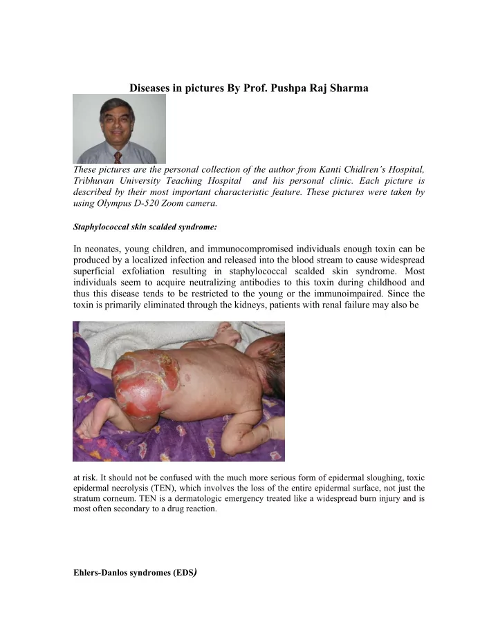

Diseases in pictures By Prof. Pushpa Raj Sharma These pictures are the personal collection of the author from Kanti Chidlren’s Hospital, Tribhuvan University Teaching Hospital and his personal clinic. Each picture is described by their most important characteristic feature. These pictures were taken by using Olympus D-520 Zoom camera. Staphylococcal skin scalded syndrome: In neonates, young children, and immunocompromised individuals enough toxin can be produced by a localized infection and released into the blood stream to cause widespread superficial exfoliation resulting in staphylococcal scalded skin syndrome. Most individuals seem to acquire neutralizing antibodies to this toxin during childhood and thus this disease tends to be restricted to the young or the immunoimpaired. Since the toxin is primarily eliminated through the kidneys, patients with renal failure may also be at risk. It should not be confused with the much more serious form of epidermal sloughing, toxic epidermal necrolysis (TEN), which involves the loss of the entire epidermal surface, not just the stratum corneum. TEN is a dermatologic emergency treated like a widespread burn injury and is most often secondary to a drug reaction. Ehlers-Danlos syndromes (EDS )
The Ehlers-Danlos syndromes (EDS) are a heterogeneous group of heritable connective tissue disorders characterized by articular hypermobility, skin extensibility and tissue fragility. Tonsiillitis The typical findings are • redder-than-normal tonsils • a yellow or white coating on the tonsils a funny-sounding voice • swollen glands in the neck • fever • • bad breath Telangiectasia Telangiectasias are found in areas of cutaneous inflammation. For example, lesions of discoid lupus frequently have telangiectasias within them. Ataxia-telangiectasia (AT) is an autosomal recessive genetic disorder characterized by cerebellar ataxia, oculocutaneous telangiectasia, and immunodeficiency .
Tetralogy of fallot A cyanotic congenital heart disease characterized by right ventricular hypertrophy, pulmonary stenosis, ventricular septal defect (VSD), and dextroposition of the aorta. o X-ray: boot-shaped (coeur en sabot) concavity of L cardiac border (no PA) � normal heart size � RVH -> elevation of apical shadow � o diminished pulmonary vascularity o aortic arch to R in 20% Achondroplasia A non-lethal type of congenital dwarfism characterized by typical skeletal dysplasias (rhizometric micromelia), a large head, and neurological manifestations. There may be recurrent and multiple fractures. Limbs: rhizometric micromelia,shortened limbs, proximal > distal shortening, elbows - lack of full
extension and supination ,legs - genu varum (bowleg), hands - trident (splayed), deviated towards ulna, short and broad . Acrodermatitis enteropathica Acrodermatitis enteropathica is a rare autosomal recessive disorder characterized by abnormalities in zinc absorption. Clinical manifestations include diarrhea, alopecia, muscle wasting, depression, irritability, and a rash involving the extremities, face, and perineum. The rash is characterized by vesicular and pustular crusting with scaling and erythema. In addition, hypopigmentation and corneal edema have been described in these patients Female pseudohermaphrodite Overall, Congenital adrenal hyperplasia (CAH) is the most frequent cause of ambiguous genitalia in the newborn, constituting approximately 60% of all intersex cases. CAH produces a female pseudohermaphrodite, which is a gonadal female with a virilized phenotype.
The basic biochemical defect is an enzymatic block that prevents sufficient cortisol production. Biofeedback via the pituitary gland causes the precursor to accumulate above the block. Clinical manifestation of CAH depends on which enzymatic defect is present. Café au lait spots Neurofibromatosis (VON RECKLINGHAUSEN'S DISEASE) 1 is characterized by cutaneous neurofibromas , pigmented lesions of the skin called cafe au lait spot s , freckling in non-sun exposed areas such as the axilla, hamartomas of the iris termed Lisch nodules, and pseudoarthrosis of the tibia. Neurofibromas are benign peripheral nerve tumors composed of proliferating Schwann cells and fibroblasts. They present as multiple, palpable, rubbery, cutaneous tumors. They are generally asymptomatic; however, if they grow in an enclosed space, e.g., the intervertebral foramen, they may produce a compressive radiculopathy or neuropathy. Aqueductal stenosis with hydrocephalus, scoliosis, short stature, hypertension, epilepsy, and mental retardation may also occur.
Cavernous haemangioma A "cavernous hemangioma" is not a hemangioma but a venous malformation in which there is a dearth of smooth muscle in the wall of a large thin venous structure lined by endothelium. These never regress spontaneously, and neither glucorticoids nor IFN- α are effective. Clubbing Hypertrophic osteoarthropathy (HOA) is characterized by clubbing of digits and, in more advanced stages, by periosteal new bone formation and synovial effusions. HOA occurs in primary and familial forms and usually begins in childhood. The secondary form of HOA is associated with intrathoracic malignancies, suppurative lung disease, congenital heart disease, and a variety of other disorders and is more common in adults. Clubbing is almost always a feature of HOA but can occur as an isolated manifestation. The presence of clubbing in isolation is generally considered to represent either an early stage or an element in the spectrum of HOA. The presence of only clubbing in a patient usually has the same clinical significance as HOA. Diaphragmatic Hernia Congenital Diaphragmatic Hernia (CDH) occurs in about one in every 2,500 live births. Absence of the diaphragm may occur on the left, right or both sides, but the left side is most common. There is a wide discrepancy between the “visible” mortality reported from children’s centers, which treat only those infants who survive gestation, birth, resuscitation, transport and often major surgery, and the true mortality, based on all prenatally diagnosed cases, which has been called the “hidden mortality” of CDH.
Exanthema subitum Exanthema subitum had been speculated to be a viral disease although its pathogen is unknown. Human herpesvirus 6 (HHV-6), first isolated in 1986, was proved by Yamanishi et al. to be the causal agent of exanthema subitum. The typical presentation is appearance of generalized macular rash when the fever subsides without other localizing signs. The common age group affected are less than two years. Caroli’s Disease Caroli disease is a nonobstructive dilatation of the intrahepatic bile ducts. This is a rare congenital disorder that classically causes saccular ductal dilatation, which usually is segmental. Caroli disease is associated with recurrent bacterial cholangitis and stone formation. Caroli disease also is known as communicating cavernous ectasia or congenital cystic dilatation of the intrahepatic biliary tree. It is distinct from other diseases that cause ductal dilatation caused by obstruction. It is not one of the many choledochal cyst derivatives.
Actinomycosis Actinomycosis is a chronic, suppurating, granulomatous condition caused by bacteria producing branching hyphae, such as Actinomyces israelii. Most commonly affecting the cheek or mandibular skin, lesions can also be located in the thorax or elsewhere. Actinomyces , produces a chronic fibrotic necrotizing process that crosses tissue planes and may involve the pleural space, ribs, vertebrae, and subcutaneous tissue, with eventual discharge of sulfur granules (macroscopic bacterial masses) through the skin (empyema necessitatis). Typically, there is a group of dull red nodules, with sinuses draining colonies of organisms called "sulfur" granules. In the mouth and elsewhere, the lesion extends from a focus of bone involvement, or from some deeper focus. Treatment: - Long term (more than a year), high dose (10 million units daily) penicillin may be required, with or without surgical debridement. - Alternatives include imipenem and erythromycin.
Recommend
More recommend