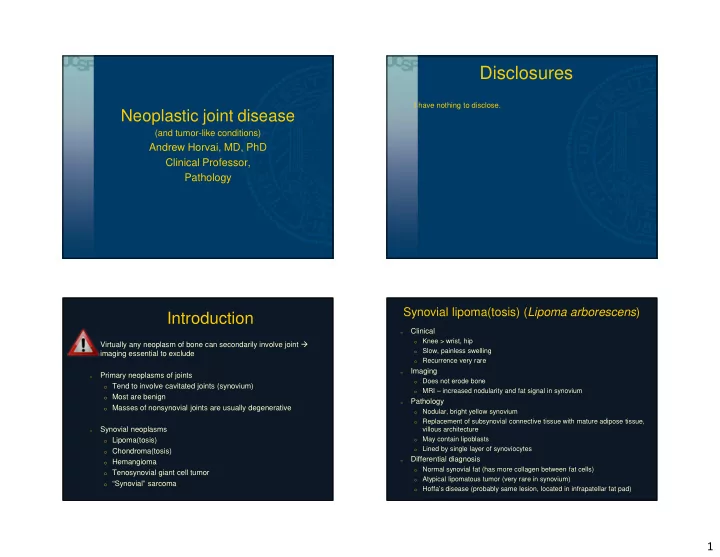

Disclosures I have nothing to disclose. Neoplastic joint disease (and tumor-like conditions) Andrew Horvai, MD, PhD Clinical Professor, Pathology Synovial lipoma(tosis) ( Lipoma arborescens ) Introduction Clinical o o Knee > wrist, hip Virtually any neoplasm of bone can secondarily involve joint � o Slow, painless swelling o imaging essential to exclude o Recurrence very rare Imaging o Primary neoplasms of joints o o Does not erode bone o Tend to involve cavitated joints (synovium) MRI – increased nodularity and fat signal in synovium o o Most are benign Pathology o o Masses of nonsynovial joints are usually degenerative o Nodular, bright yellow synovium o Replacement of subsynovial connective tissue with mature adipose tissue, Synovial neoplasms villous architecture o May contain lipoblasts o Lipoma(tosis) o o Lined by single layer of synoviocytes o Chondroma(tosis) Differential diagnosis o o Hemangioma o Normal synovial fat (has more collagen between fat cells) o Tenosynovial giant cell tumor Atypical lipomatous tumor (very rare in synovium) o o “Synovial” sarcoma o Hoffa’s disease (probably same lesion, located in infrapatellar fat pad) 1
Synovial lipoma Synovial lipomatosis Does not erode bone T2 FS 1 cm Synovial lipomatosis Synovial lipomatosis 2
Synovial chondromatosis Synovial chondroma(tosis) Clinical o 3 rd -5 th decade o o Knee, shoulder, large joints Rare recurrence o o Malignant transformation ~1% Imaging o o If calcified, plain film shows multiple cloudy or ring calcs Can erode cortex o o MRI, lobulated, bright on T2 Pathology o o Multiple nodules of blue cartilage, + lined by synovium Clustering of chondrocytes o o Nucleomegaly and binucleation common, mitoses absent o Necrosis OK Differential diagnosis o o Chondro-osseous loose body o Soft tissue chondroma (closely related if not same) o Synovial chondrosarcoma (secondary chondrosarcoma) Synovial chondromatosis: multiple nodules, eroding articular cartilage Synovial chondromatosis: clustering + endochondral ossification 3
Synovial chondromatosis Synovial chondromatosis Synovial chondromatosis Synovial chondromatosis, secondary chondrosarcoma 4
Chondro-osseous loose body Synovial chondromatosis, secondary chondrosarcoma Chondro-osseous loose body: Chondro-osseous loose body Hyaline and fibrocartilage, irregular calcifications 5
Synovial “hemangioma” Synovial “hemangioma” Clinical o Vascular malformation not a true vascular neoplasm o o Children > Adults, knee most common o Most asymptomatic, or slow swelling, decreased range of motion o Cured by excision Imaging o o Plain film may suggest effusion (nonspecific) o MRI – T2 bright serpentine structures, fluid-fluid levels Pathology o Vascular channels of varying caliber, increased density beneath normal or o hyperplastic synovium o Increased fat o No atypia or mitoses Differential diagnosis o Chronic hemarthrosis (grossly) o o Diffuse tenosynovial giant cell tumor (imaging) T2 FS Synovial hemangioma Synovial “hemangioma” 6
Synovial hemangioma Synovial hemangioma Tenosynovial giant cell tumor Tenosynovial giant cell tumor Clinical o o Adults, knee, wrist, digits, shoulder Localized Diffuse Slow growing joint swelling, locking o o Recurrence risk ~ growth pattern Gender M > F F > M Imaging o Joints Small, hand Large, knee o Soft tissue mass, may erode bone “Bloom” on MRI, T2 bright, sometimes T1 bright also o Size < 5 cm >10 cm Pathology o Recurrence 4-30% 30-50% o Polymorphic: Histiocytes, multinucleated giant cells, spindly fibroblasts, siderophages, foam cells, chronic inflammation IHC CD68, desmin CD68, desmin o Central fibrosis in older lesions Mitoses may be brisk not atypical o Differential diagnosis o o Giant cell tumor of low malignant potential (soft tissue analog of GCT of bone) o Chronic hemarthrosis Malignant tenosynovial giant cell tumor o 7
Tenosynovial giant cell tumor: Diffuse Tenosynovial giant cell tumor Diffuse, T2 FS Localized, T2 FS Horvai A, Robbins Basic Pathology, Elsevier, 2017 Tenosynovial giant cell tumor: Diffuse Tenosynovial giant cell tumor: Diffuse 8
Tenosynovial giant cell tumor: diffuse Tenosynovial giant cell tumor: diffuse Tensynovial giant cell tumor: Tenosynovial giant cell tumor: diffuse Tenosynovial giant cell tumor: localized localized 9
Tenosynovial giant cell tumor: localized Tenosynovial giant cell tumor: localized Tenosynovial giant cell tumor: localized Tenosynovial giant cell tumor: localized 10
Tenosynovial giant cell tumor: localized Tenosynovial giant cell tumor: localized Tenosynovial giant cell tumor: desmin Tenosynovial giant cell tumor Same genetic abnormality in both subtypes ( COL6A3:CSF1 ) � Diffuse and localized type related to anatomic compartment � rather than biology Provides mechanism of pathogenesis analogous to giant cell � tumor of bone West RB et al. PNAS, 2006 103(3) 690–695. 11
Synovial sarcoma Synovial sarcoma Clinical o o Young adults (peak), wide age range o 80% extremity, knee common but not intra-articular, can arise anywhere o 5 year survival 36-76%, chemotherapy + surgery Pathology o Monophasic (70%): uniform spindle cells, short fascicles, herringbone o Biphasic (30%): uniform spindle cells + pseudoglands o Jones SF, Whitman RC. Ann Surg. 1914;60(4):440-50. Poorly differentiated (<1%): small round blue cell tumor o o Hyperchromatic nuclei, paradoxically low mitoses o Branching vessels, calcification IHC o o Keratin, EMA – usually focal and patchy o S100 in ~30%, SOX10 <10% (usually intraneural synovial sarcoma) o CD34 negative Genetics o t(X;18), SS18-SSX1 –SSX2, -SSX4 fusion o Monophasic synovial sarcoma Monophasic synovial sarcoma 12
Monophasic synovial sarcoma Monophasic synovial sarcoma Biphasic synovial sarcoma Biphasic synovial sarcoma 13
Poorly differentiated synovial sarcoma Monophasic synovial sarcoma Keratin Keratin Take-home messages 1. Do not diagnose diseases of joints (especially synovial chondroma(tosis) without reviewing imaging. It could be a bone tumor. 2. Just because it’s villous and synovial it does not mean it is Pigmented Villonodular Synovitis 3. Do not over-diagnose a desmin positive PVNS as EMA SS18 break apart FISH rhabdomyosarcoma 4. Synovial sarcoma is a sarcoma but has nothing to do with normal synovial cells 14
Recommend
More recommend