Disclosures Joo A.C. Lima, M.D. Receives grant /research support - PowerPoint PPT Presentation
Disclosures Joo A.C. Lima, M.D. Receives grant /research support from Toshiba Medical Systems Bracco Diagnostics Federal Grant Support: NHLBI, NIDDK, and NIA Diagnostic Performance of Combined Noninvasive Coronary Angiography
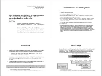
Disclosures João A.C. Lima, M.D. • Receives grant /research support from – Toshiba Medical Systems – Bracco Diagnostics – Federal Grant Support: NHLBI, NIDDK, and NIA
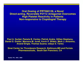
Diagnostic Performance of Combined Noninvasive Coronary Angiography and Myocardial Perfusion Imaging Using 320-row Detector Computed Tomography: The CORE320 Multicenter International Study João A.C. Lima, M.D., Johns Hopkins Hospital Background • The benefits of revascularization are highest in patients who have coronary stenoses that are flow limiting and hemodynamically significant. • Invasive angiography and CT angiography are limited in delineating flow- limiting lesions which are detected by perfusion imaging or invasive FFR. • A single test which can non invasively evaluate the severity of a lesion and the hemodynamic significance is desirable for the management of patients with symptomatic CAD.
Main Objectives/Study Design To evaluate: • The diagnostic performance of combined CTA and CTP to identify patients with flow limiting CAD compared with invasive angiography and SPECT-MPI • Incremental value of CTP above CTA alone • Prediction of coronary revascularization vs. ICA + SPECT • 381 patients from 16 hospitals in 8 countries who were clinically referred for ICA underwent SPECT-MPI and a combined CT angiography and myocardial perfusion scan.
Core Laboratory Image Analysis Coronary Image Analysis Myocardial Perfusion Analysis Angiography Core Lab SPECT-MPI Core Lab CT Angiography Core Lab CT Perfusion Core Lab Two readers Two readers • Double Blinded Analysis • Differences resolved by consensus • Differences resolved by consensus Entire coronary tree analyzed 13 Segment myocardial model • 19 segment model • Visual assessment • Stented segments included 0 = normal • Visual assessment on all segments 1 = mild perfusion deficit • Stenosis > 30% quantified 2 = moderate perfusion deficit • Maximum % stenosis 3 = severe perfusion deficit
Baseline Characteristics Age – Median [IQR] 62 [56-68] Men – number [%] 258 [66%] Body Mass Index – Median [IQR] 27 [24-30] Hypertension – number [%] 302 [78%] Diabetes – number [%] 132 [34%] Dislipidemia – number [%] 261 [68%] Previous MI – number [%] 95 [25%] Smoking (Current + Former) – number [%] 202 [53%] Prior PCI – number [%] 112 [29%] Family history of CAD – number [%] 167 [45] Creatinine – mg/dl – Median [IQR] 0.9 (0.7-1.0)
Results Incremental Value of CTA-CTP over CTA CTA-CTP vs. ICA/SPECT to predict (Reference Standard: 50% by ICA with Vessel Level Revascularization SPECT-MPI defect) (Reference Standard: Revascularization at 30 days) 1.0 1.0 0.9 0.9 P<0.001 0.8 0.8 P = 0.35 0.7 0.7 Sensitivity Sensitivity 0.6 0.6 0.5 0.5 CTA-CTP ROC Area = 0.79 CTA-CTP ROC Area = 0.87 0.4 0.4 95% CI [0.76-0.83] 95% CI [0.83-0.91] 0.3 0.3 ICA-SPECT ROC Area = 0.81 CTA ROC Area = 0.81 95% CI [0.78-0.84] 0.2 0.2 95% CI [0.77-0.86] 0.1 0.1 N=381 QCA+SPECT CTA+CTP CT Prevalence = 38% CTA alone 0.0 0.0 0.0 0.1 0.2 0.3 0.4 0.5 0.6 0.7 0.8 0.9 1.0 0.0 0.1 0.2 0.3 0.4 0.5 0.6 0.7 0.8 0.9 1.0 1-Specificity 1-Specificity
Patient Based Results – Known CAD Excluded Patient-Based Analysis for participants without history of CAD (Reference Standard: 50% by ICA with a corresponding myocardial perfusion defect on SPECT-MPI) 1.0 0.9 0.8 Patient Based Combined CTA-CTP 0.7 vs. Sensitivity 0.6 Reference Standard (ICA 50% with SPECT- ROC area = 0.93 0.5 MPI defect) 95% CI [0.89-0.96] 0.4 0.3 0.2 0.1 N=231 Prevalence = 26% 0.0 0.0 0.1 0.2 0.3 0.4 0.5 0.6 0.7 0.8 0.9 1.0 1-Specificity
Patients with Known CAD Excluded Sensitivity Specificity PPV NPV CTA alone 93 60 46 96 ≥ 50% Stenosis (95% CI) (84-98) (52-67) (37-55) (91-99) CTP SSS 97 58 45 98 0 (89-100) (50-65) (36-54) (93-100) 90 67 49 95 1 (80-96) (59-74) (40-59) (89-98) 89 69 51 94 2 (78-95) (61-76) (41-60) (89-98) 84 74 54 93 3 (72-92) (67-81) (43-64) (87-96) 80 80 59 92 4 (68-89) (73-86) (48-70) (86-96) 71 87 65 89 5 (57-82) (80-91) (52-77) (83-93)
Conclusions • Combined CTA-CTP can detect flow-limiting stenoses defined by ICA (50% or greater) with an associated SPECT- MPI defect. • CT perfusion adds significantly to the diagnostic power of CT angiography alone. • The combination of CTA & CTP in one non-invasive exam is useful in identifying the patients who will benefit the most from revascularization and to guide the management of CAD.
Recommend
More recommend
Explore More Topics
Stay informed with curated content and fresh updates.
