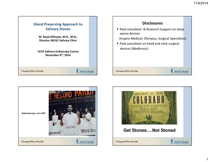

11/6/2014 Disclosures Gland Preserving Approach to Salivary Stones � Paid consultant & Research Support on sleep apnea devices M. Boyd Gillespie, M.D., M.Sc. (Inspire Medical; Olympus; Surgical Specialties) Director, MUSC Salivary Clinic � Paid consultant on head and neck surgical devices (Medtronic) UCSF Salivary Endoscopy Course November 6 th , 2014 Sialendoscopy circa 2007 Get Stones….Not Stoned 1
11/6/2014 Introduction: Salivary Stones The Limitations of Surgery � Major source of obstructive salivary swelling � Est. incidence 1 in 10,000-20,000 per year � Risk factor- smoking; dry mouth (medications) � Mucous core surrounded by inorganic shell (calcium hydroxylapatite); Mean growth 1mm/year � Readily diagnosed by US or CT (contrast not needed) � Stones < 2mm may be missed by either technique High-rate of FN paresis (40-50%) and paralysis (5%) after gland resection for � inflammatory disorders. Glands with chronic sialadenitis are often histologically normal � Marchal F, et al. Ann Otol Rhinol Laryngol. 2001; 110: 464-469. Glands with chronic sialadenitis can resume normal function if the � obstruction is relieved. Su YX, et al. Laryngoscope 2009; 119: 464-452. Ambulatory Operative � Procedure Treatment strategy General Anesthesia � I. Small Stones/Mobile Stones (1-5mm) Nasal Intubation (SMG) Interventional Endoscopy � Oral Intubation (Parotid) � � OR time 2 hours � II. Medium sized stones (5-10mm) Endoscopy + combined measures � III. Large Stones (>10mm) Combined approaches � IV. Gland removal Multiple stones (>3) or failures 2
11/6/2014 Small Stones Interventional Sialendoscopy Case 1 for treatment Small ( ≤ 5 mm) Mobile Salivary Stones (Parotid or SMG) 0.38-0.6 mm 0.78 mm 0.38-0.78 mm � 23 year old male presents with intermittent left parotid gland swelling. � CT scan suggests a small 3mm stone at the hilum of the parotid. � Patient continues to have symptoms after 0.7 mm several weeks of hydration, massage, and sialogogues. 9 1 st line- Endoscopic Basket Retrieval 3 mm stone at hilum of left parotid 3
11/6/2014 5 mm stone at hilum of left parotid Case 2 Small ( ≤ 5 mm) Fixed Salivary Stones (Parotid or SMG) � 46 year old woman presents with intermittent left parotid gland swelling. � CT scan suggests a 5 mm stone at of the hilum of the left parotid. � Patient continues to have symptoms after several weeks of hydration, massage, and sialogogues. 1 st Line- Intermediate Stones Endoscopic Approach with Stone Shattering Case 3 Intermediate (5-10 mm) or Fixed Salivary Stones � 55 year old woman presents with intermittent right SMG swelling. � CT scan suggests a 8 mm stone at proximal right Wharton’s duct. � Patient has had several cases of acute sialadenitis in last 3 months requiring 2 course of antibiotics. 4
11/6/2014 1 st Line- Endoscopic Shattering versus 8 mm stone in proximal Wharton’s duct Combined Approach (Endoscopic-Open) Technique � Step 1- Insert scope to confirm stone is not amendable to endoscopic shattering (hard, fixed, too large); Irrigation of infection and debris; Dilation of duct (assist with visualization during open approach). � Step 2- Incision of tissue overlying duct/ gland. � Step 3- Stone localization with endoscope. � Step 4- Direct opening of duct with stone extraction. � Step 5- Passage of scope to irrigate gland and remove remaining fragments. � Step 6- Repair duct (Sialodochoplasty-SMG) Step 1- Determine if Amendable to Endoscopic Shaterring (Forceps; Hand Drill; Laser) Endoscopic Removal Intracorporeal Laser Lithotripsy (Holmium; 200 micron; 2.5-3.5 watts) 5
11/6/2014 Step 2- Plan Incision Step 4- Opening of duct with direct stone extraction Case 4 Step 4- Opening of duct with direct stone extraction Intermediate (5-10 mm), or Fixed Parotid Salivary Stones � 52 year old man presents with intermittent left parotid gland swelling. � US of gland: Lymph node v. stone? � 6 mm irregular stone found on diagnostic sialendoscopy 6
11/6/2014 Intermediate Stone (5-10mm): 1st Line: Endoscopic Shaterring Endoscopic Mobilization and Shattering (Basket, Handrill, Laser, Forceps) Papillotomy (Basket; Forceps; Hand Drill; Laser) Video Courtesy of Johannes Zenk, MD, PhD 26 Case 5 Papillotomy Intermediate to Large (> 5 mm), Deep (beyond scope), or Fixed Parotid Salivary Stones � 36 year old man presents with intermittent right parotid gland swelling. � CT scan suggests a 8 mm stone at hilum of gland. 7
11/6/2014 1 st Line- Parotid Transfacial Approach (Endoscopic-Open) 8mm stone at hilum of right parotid Technique � Step 1- Apply NIMS; Insert scope to confirm stone is not amendable to endoscopic shattering (hard, fixed, too large); Irrigation of infection and debris; Dilation of duct (assist with visualization during open approach). � Step 2- Raise preauricular flap � Step 3- Stone localization with endoscope/ US (needle). � Step 4- Divide parotid fascia and gland � Step 5- Direct opening of duct with stone extraction. � Step 6- Passage of scope to irrigate gland and remove remaining fragments. � Step 7- Repair duct (5.0 PDS) and close fascia; pressure dressing. NIMS on buccal branch Endoscopic Localization 8
11/6/2014 Localization of Stone: Endoscopy Localization of Stone: Ultrasound 23 Gauge Needle with Methylene Blue Opening of duct with direct stone extraction Pass scope again to remove remaining fragments 9
11/6/2014 Repair Duct Transfacial Stone Removal 5.0 PDS for duct and 4.0 vicryl for fascia. Apply jaw bra pressure dressing for 72 hours. Case 5 Intermediate to Large (> 5 mm) or Fixed Stones within 2 cm of Parotid Ostium Mean follow-up of 1 year � 10/14 (71%) stone-free and symptom-free � 3/14 (21%) stone-free and improved with intermittent symptoms � 1/14 (7%) required follow-up parotidectomy � Complications in 4/14 (29%)- 2 with periauricular anesthesia, � 1 salivary fistula, 1 sialocele. Facial nerve seen in 5/14 (36%) of cases � 10
11/6/2014 1 st Line- Parotid Transbuccal Approach (Endoscopic-Open) 5mm stone fixed 1cm beyond left parotid ostium Technique � Step 1- Insert scope to confirm stone is not amendable to endoscopic shattering (hard, fixed, too large); Irrigation of infection and debris; Dilation of duct (assist with visualization during open approach). � Step 2- Semilunar incision anterior to ostium; divide buccinator fibers. � Step 3- Localize stone with endoscopic transillumination. � Step 4- Open duct and remove stone. � Step 5- Repair duct (5.0 PDS) and close mucosa. � Step 6- Consider ductal stent (Hood; Sialotechology) Transbuccal Incision Repair Duct 11
11/6/2014 Consider Stent Direct Transfacial Approach Massive Parotid Stone with Abscesses Gland Excision Massive Parotid Stone with Abscesses Questions or Concerns: gillesmb@musc.edu 12
Recommend
More recommend