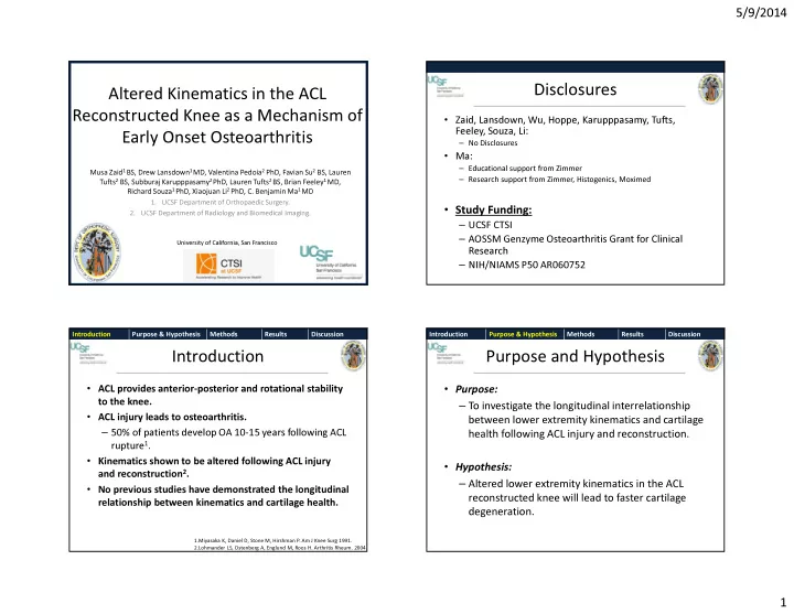

5/9/2014 Disclosures Altered Kinematics in the ACL Reconstructed Knee as a Mechanism of • Zaid, Lansdown, Wu, Hoppe, Karupppasamy, Tufts, Feeley, Souza, Li: Early Onset Osteoarthritis – No Disclosures • Ma: – Educational support from Zimmer Musa Zaid 1 BS, Drew Lansdown 1 MD, Valentina Pedoia 2 PhD, Favian Su 2 BS, Lauren – Research support from Zimmer, Histogenics, Moximed Tufts 2 BS, Subburaj Karupppasamy 2 PhD, Lauren Tufts 2 BS, Brian Feeley 1 MD, Richard Souza 1 PhD, Xiaojuan Li 2 PhD, C. Benjamin Ma 1 MD 1. UCSF Department of Orthopaedic Surgery. • Study Funding: 2. UCSF Department of Radiology and Biomedical Imaging. – UCSF CTSI – AOSSM Genzyme Osteoarthritis Grant for Clinical University of California, San Francisco Research – NIH/NIAMS P50 AR060752 Introduction Purpose & Hypothesis Methods Results Discussion Introduction Purpose & Hypothesis Methods Results Discussion Introduction Purpose and Hypothesis • ACL provides anterior-posterior and rotational stability • Purpose: – To investigate the longitudinal interrelationship to the knee. • ACL injury leads to osteoarthritis. between lower extremity kinematics and cartilage – 50% of patients develop OA 10-15 years following ACL health following ACL injury and reconstruction. rupture 1 . • Kinematics shown to be altered following ACL injury • Hypothesis: and reconstruction 2 . – Altered lower extremity kinematics in the ACL • No previous studies have demonstrated the longitudinal reconstructed knee will lead to faster cartilage relationship between kinematics and cartilage health. degeneration. 1.Miyasaka K, Daniel D, Stone M, Hirshman P. Am J Knee Surg 1991. 2.Lohmander LS, Ostenberg A, Englund M, Roos H. Arthritis Rheum. 2004. 1
5/9/2014 Introduction Purpose & Hypothesis Methods Results Discussion Introduction Purpose & Hypothesis Methods Results Discussion Methods Methods • Subjects: • Kinematic MRI: N=20 – Use MRI to determine 3-dimensional Average Age 29 +/- 8.9 years knee kinematics 3 . 23.2 +/- 3.1 kg/m 2 Average BMI – Sagittal T2 FSE images obtained in Gender 10 males, 10 females extension and knee flexion loaded with • 25% of total body weight. Exclusion Criteria: – Other ligamentous injuries, meniscal injury requiring repair, previous – Customized in house software used to history of arthritis, knee trauma, or surgery. determine tibial and femoral axis 3 . – Determine position of tibia relative to ACL femur in the extended and flexed Baseline 1 Year MRI ACL Rupture 6 Month MRI Reconstruction MRI states. 3. Carpenter R, Majumdar S, Ma C. Arthroscopy 2009 Introduction Purpose & Hypothesis Methods Results Discussion Introduction Purpose & Hypothesis Methods Results Discussion Methods Average Tibial Position in Knee Extension • 25 Cartilage T1p MRI: * Posterior – Quantifies proteoglycan content 4 . – Detection * 20 of early cartilage damage 4 . – Comprehensive quantification of 15 Avg Ipsi Extended Tibial cartilage T1p relaxation times of the Position medial and lateral femoral condyles Avg Contra Extended Tibial 10 Position and tibial cartilage was measured with customized software. * Indicates p < 0.05 5 – Cartilage Anterior was divided into subcompartments to examine 0 regional variations in cartilage Baseline 1 Year damage. Tibial position is shifted more anterior relative to the femur in the extended state at baseline. 4. Li X, Ma C, Link T, et al. Osteoarthritis and Cartilage. 2007 2
5/9/2014 Introduction Purpose & Hypothesis Methods Results Discussion Introduction Purpose & Hypothesis Methods Results Discussion Side to Side Difference in Extended Tibial Position vs Average Tibial Position in Knee Flexion Progression of T1p in LFC from Baseline to 1 Year 8 * 25 Posterior Progression of T1p in the LFC from 6 Baseline to 1 Year 4 20 * 2 15 0 Avg Ipsi Flexed Tibial Position -3 -2 -1 0 1 2 3 4 -2 Avg Contra Flexed Tibial 10 Spearman’s Rho = 0.44 Position -4 p = 0.046 * Indicates p < 0.05 -6 5 SSD in Tibial Position in the Extended State at 1 Year Anterior 0 The side to side difference in the extended tibial position Baseline 1 Year correlates to the progression of cartilage T1p in the lateral Tibial position is shifted more anterior relative to the femur femoral condyle. in the flexed state at baseline. Introduction Purpose & Hypothesis Methods Results Discussion Introduction Purpose & Hypothesis Methods Results Discussion Side to Side Difference in Flexed Tibial Position vs Discussion Progression of T1p in pLT from Baseline to 1 Year 15 • Knee kinematic are altered following ACL injury. Progression of T1p in the Posterior LT – Tibia more anterior in extended and flexed states at baseline. 10 from Baseline to 1 Year – Normalization of kinematics 1 year post reconstruction. 5 • Alterations in knee kinematics are related to detectable 0 cartilage degeneration in the knee as soon as 1 year -6 -4 -2 0 2 4 -5 following ACL reconstruction. • Longitudinal relationship between kinematics and the -10 Spearman’s Rho = 0.444 P = 0.044 progression of cartilage damage. -15 – Alterations in knee kinematics at 1 year post reconstruction are correlated to SSD in the Flexed Tibial Position at 1 Year the progression of T1p from baseline to 1 year. The side to side difference in the flexed tibial position correlates to the progression of cartilage T1p in the posterior lateral tibia from baseline to 1 year. 3
5/9/2014 Introduction Purpose & Hypothesis Methods Results Discussion Discussion Acknowledgements • • Haughom et al has demonstrated a relationship between C. Benjamin Ma, MD • Xiaojuan Li, PhD • Brian Feeley, MD altered knee kinematics and cartilage damage 4 . • Richard Souza, PT PhD • • Our study is unique as it follows patients longitudinally to Sharmila Majumdar, PhD • Christina Allen, MD • Drew Lansdown, MD assess how altered kinematics relate to cartilage damage • Valentina Pedoia, PhD • Favian Su, BS over time. • Lauren Tufts, BS • • Understanding the longitudinal interrelationship between Subburaj Karupppusamy, PhD • UCSF Department of Orthopaedic Surgery • Musculoskeletal Quantitative Imaging Group the kinematics in the ACL reconstructed knee and signs of • UCSF Department of Radiology • The CTRFP Team early cartilage change may provide insight into the development and prevention of post-traumatic Funding: • AOSSM Genzyme Osteoarthritis Grant for Clinical Research osteoarthritis. • NIH/NIAMS P50 AR060752 • Will continue longitudinal follow up at 2 and 3 years. • CTSI/UCSF Clinical and Translational Research Fellowship 4. . Haughom B, et al, The Knee, August 2012, 482-487 4
Recommend
More recommend