SLIDE 1 Communication Between Neurons
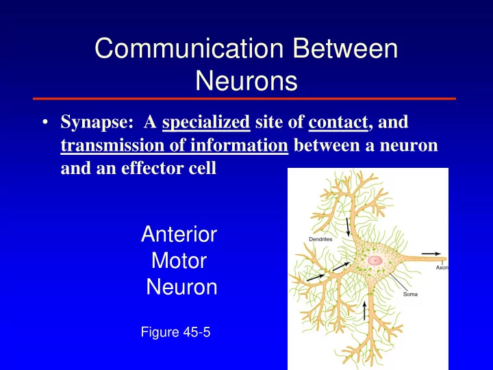
- Synapse: A specialized site of contact, and
transmission of information between a neuron and an effector cell
Figure 45-5
Anterior Motor Neuron
SLIDE 2 Communication Between Neurons
Neurotransmitter: is a messenger of neurologic information from
SLIDE 3 Action of Neurotransmitter on Postsynaptic Neuron
- postsynaptic membrane contains receptor
proteins for the transmitter released from the presynaptic terminal.
- The effect of neurotransmitter on the post
synaptic neuron depend on the type of the receptor
SLIDE 4 Action of Neurotransmitter on Postsynaptic Neuron
– Ion channels receptors
SLIDE 5 Action of Neurotransmitter on Postsynaptic Neuron
– Ion channels receptors Ionotropic – Second messenger receptors Metabotropic
SLIDE 6 Ion Channels receptors
- transmitters that open sodium
channels excite the postsynaptic neuron.
- transmitters that open chloride
channels inhibit the postsynaptic neuron.
- transmitters that open potassium
channels inhibit the postsynaptic neuron.
SLIDE 7 Seconded messenger receptors (as example G-protein)
Ion Channel
channels
cGMP
more intracellular enzymes
transcription.
SLIDE 8 G-Protein-Coupled Receptors and Effectors
- GPCR Effector Systems (Cont’d)
- Push-pull method (e.g., different G proteins for
stimulating or inhibiting adenylyl cyclase)
SLIDE 9 G-Protein-Coupled Receptors and Effectors
- GPCR Effector Systems (Cont’d)
- Some cascades split
– G-protein activates PLC→ generates DAG and IP3→ activate different effectors
SLIDE 10 G-Protein-Coupled Receptors and Effectors
(Cont’d)
SLIDE 11 Drugs and the Synapse 1) at the receptor
- The study of the influence of various kinds of drugs has
provided us with knowledge about many aspects of neural communication at the synaptic level.
- Drugs either facilitate or inhibit activity at the synapse.
– Antagonistic drugs block the effects of neurotransmitters (e.g., novacaine, caffeine). – Agonist drugs mimic or increase the effects of neurotransmitters (e.g., receptors in the brain respond to heroin, LSD and cocaine) – Allosteric modulation
SLIDE 12 Drugs and the Synapse
- A drug has an affinity for a particular type of
receptor if it binds to that receptor.
– Can vary from strong to weak.
- The efficacy of the drug is its tendency to activate
the receptor .
- Drugs can have a high affinity but low efficacy.
SLIDE 13
Agonists and Antagonists
SLIDE 14
Agonists and Antagonists
SLIDE 15
Agonists and Antagonists
SLIDE 16
Agonists and Antagonists
SLIDE 17
Allosteric modulation
SLIDE 18
Synaptic Transmission
SLIDE 19 Drugs and the Synapse 2) alter various stages of synaptic processing.
- Drugs work by doing one or more of the
following to neurotransmitters:
1. Increasing the synthesis. 2. Causing vesicles to leak. 3. Increasing release. 4. Decreasing reuptake. 5. Blocking the breakdown into inactive chemical. 6. Directly stimulating or blocking postsynaptic receptors.
SLIDE 20 Neurotransmitters
- Synthesis : esp. rate-limiting enzyme and/or
substrate
- Clearance and inactivation
- Location and pathway
- Dysfunction and CNS pathology
SLIDE 21 Neurotransmitters
- More than 50 chemical substances does
function as synaptic transmitters.
– small molecules which act as rapidly acting transmitters.
- acetylcholine, norepinephrine, dopamine,
serotonin, GABA, glycine, glutamate, NO. – neuropeptides.
- endorphins, enkephalins, VIP, ect.
- hypothalamic releasing hormones.
– TRH, LHRH, ect.
– ACTH, prolactin, vasopressin, ect.
SLIDE 22
Fast Neurotransmitteres
SLIDE 23 Glutamate (L-glutamic acid)
- Main excitatory neurotransmitter in the
mammalian CNS
- 95% of excitatory synapses in the brain are
glutamatergic
- Precursor for the GABA (major inhibitory
neurotransmitter)
SLIDE 24 Enzymatic Pathways Involved in the Metabolism
Glutamate
Gluck et al, Am J Psychiatry 2002; 159;1165-1173
SLIDE 25 Slow synaptic transmission Fast synaptic transmission
SLIDE 26 NMDA AMPA Kainate
Kainate
Na+ Ca++ presynaptic postsynaptic 95% of excitatory synapses in the brain are glutamatergic
Kainate
SLIDE 27 The Glutamate Synapse
Note – significant Glu uptake (mainly astrocytes) Interconversion of glutamate to glutamine
SLIDE 28
Glutamate and CNS disorders
1) Stroke Ischemia →
SLIDE 29
Glutamate and CNS disorders
1) Stroke Ischemia → no ATP →
SLIDE 30
Glutamate and CNS disorders
1) Stroke Ischemia → no ATP → increase Glutamate →
SLIDE 31
Glutamate and CNS disorders
1) Stroke Ischemia → no ATP → increase Glutamate → Over activation NMDA R & AMPA R →
SLIDE 32
Glutamate and CNS disorders
1) Stroke Ischemia → no ATP → increase Glutamate → Over activation NMDA R & AMPA R → increase Ca+ → cell death 2) dysfunction of glutamatergic transmission may also involve in schizophrenia-like symptoms, cognitive dysfunction, Depression and memory impairment
SLIDE 33 GABA
- Main inhibitory neurotransmitter in the
mammalian CNS
SLIDE 34 GABA
- Main inhibitory neurotransmitter in the
mammalian CNS Ionotropic
GABAA Heterooligomeric protein complex that consists of several binding sites coupled to an integral Cl- channel
Metabotropic
GABAB G - protein coupled receptor, seven transmembrane domain protein
SLIDE 35 GABA-A- ionotropic receptor
- An integral chloride channel activated by competitive agonists: GABA
and muscimol
- Blocked by convulsant bicuculine (competitive antagonist) and
picrotoxin (noncompetitive antagonist)
- Allosterically modulated by benzodiazepines and barbiturates,
which potentiate the effect of GABA
SLIDE 36 GABAA receptor
Actions at GABAA Receptors
SLIDE 37 GABA A and ethanol
⚫ Ethanol facilitates GABA ability to activate the
receptor and prolongs the time that the Cl- channel remains open
SLIDE 38
GABA
Glutamate
GABA
GAD
GABA is formed by the α-decarboxylation of glutamate in the reaction catalyzed by GAD (glutamic acid decarboxylase)
Synthesis
SLIDE 39
GABA
GABA
GABA-T
succinic semialdehyde
GABA is catabolized into the succinic semialdehade in the reaction catalyzed by GABA-T (GABA-Transaminase)
Degradation
SLIDE 40
SLIDE 41
EEG and Seizures
SLIDE 42 Seizure Pathophysiology
- Altered ionic conductance (increased excitability)
- f neuron.
- Reduced inhibitory neuronal (primarily
GABAergic) control.
- Increased excitatory neuronal (primarily
glutamatergic) control.
- Probable mechanisms tend to overlap.
SLIDE 43
Neuromodulators
SLIDE 44 Acetylcholine
Choline + Acetyl CoA Acetyl choline + CoA ChAT
SLIDE 45
Acetylcholine synapse
SLIDE 46
Acetylcholine receptors
SLIDE 47 Acetylcholine Pathway
Nucleus basalis
SLIDE 48 Acetylcholine Pathway
Nucleus basalis
- arousal and sleep wake cycle
- enhancement of sensory
perceptions
- sustaining attention
- reward
SLIDE 49 Acetylcholine Pathway
Nucleus basalis
- arousal and reward
- enhancement of sensory
perceptions
Alzheimer’s disease – loss of cholinergic cells in nucleus basalis
SLIDE 50
Biogenic Amines
SLIDE 51 08/20/2008 Lerant: Catecholamines 2008 51
The biosynthetic pathway for the catecholamine neurotransmitters
SLIDE 52
Biogenic Amines Synapses MAO : Monoamine Oxidase
SLIDE 53
Dopamine
SLIDE 54 Dopamine receptors
- G protein-coupled receptors
SLIDE 55 Dopamine receptors
- G protein-coupled receptors
- D1 → excite
- D2 → inhibit
- D3 → inhibit
- D4 → inhibit
- D5 → excite
SLIDE 56 Dopamine receptors
- G protein-coupled receptors
- D1 → excite
- D2 → inhibit Mainly presynabtic (Autoreceptor)
- D3 → inhibit
- D4 → inhibit
- D5 → excite
SLIDE 57 08/20/2008 Lerant: Catecholamines 2008 57
(DA) synapse
SLIDE 58
Dopamine Pathways
SLIDE 59 Lerant: Catecholamines 2008
DOPAMINERGIC PATHWAYS
Substrantia nigra
Ventral tegmental area
Striatum Nigrostriatal pathway Nucl. accumbens Mesolimbic pathway Prefrontal CTX Mesocortical pathway
SLIDE 60 Lerant: Catecholamines 2008
DOPAMINERGIC PATHWAYS
Substrantia nigra
Ventral tegmental area
Striatum Nigrostriatal pathway Nucl. accumbens Mesolimbic pathway Prefrontal CTX Mesocortical pathway
- Degeneration of nigro-striatal DA system
and Decreased DAergic trans-mission in the basal ganglia will lead to
SLIDE 61 Lerant: Catecholamines 2008
DOPAMINERGIC PATHWAYS
Substrantia nigra
Ventral tegmental area
Striatum Nigrostriatal pathway Nucl. accumbens Mesolimbic pathway Prefrontal CTX Mesocortical pathway
- Degeneration of nigro-striatal DA system
and Decreased DAergic trans-mission in the basal ganglia will lead to
Parkinson Disease
SLIDE 62 Lerant: Catecholamines 2008
DOPAMINERGIC PATHWAYS
Substrantia nigra
Ventral tegmental area
Striatum Nigrostriatal pathway Nucl. accumbens Mesolimbic pathway Prefrontal CTX Mesocortical pathway
PLEASURE, REWARD AND BEHAVIOR REINFORCING PATHWAY
PLEASURE, REWARD AND BEHAVIOR REINFORCING PATHWAY
SLIDE 63 Lerant: Catecholamines 2008
DOPAMINERGIC PATHWAYS
Substrantia nigra
Ventral tegmental area
Striatum Nigrostriatal pathway Nucl. accumbens Mesolimbic pathway Prefrontal CTX Mesocortical pathway
PLEASURE, REWARD AND BEHAVIOR REINFORCING PATHWAY
natural drug-induced cocaine
SLIDE 64
SLIDE 65 Lerant: Catecholamines 2008
DOPAMINERGIC PATHWAYS
Substrantia nigra
Ventral tegmental area
Striatum Nigrostriatal pathway Nucl. accumbens Mesolimbic pathway Prefrontal CTX Mesocortical pathway
PLEASURE, REWARD AND BEHAVIOR REINFORCING PATHWAY
natural drug-induced cocaine
Hyperactivity of mesolimbic pathway:
- positive symptoms of schizophrenia
(hallucinations, etc)
SLIDE 66 Lerant: Catecholamines 2008
DOPAMINERGIC PATHWAYS
Substrantia nigra
Ventral tegmental area
Striatum Nigrostriatal pathway Nucl. accumbens Mesolimbic pathway Prefrontal CTX Mesocortical pathway
PATHWAY INVOLVED IN MOTIVATION TO EXPLORE THE ENVIRONMENT: CURIOSITY, INTEREST, COGNITIVE FLEXIBILITY, DRIVE FOR SOCIAL ENGAGEMENT. Relative hypofunction in schizophrenia: Primary mesocortical dopamine deficiency will increase the NEGATIVE SYMPTOMS like Cognitive blunting, social isolation, apathy, anhedonia