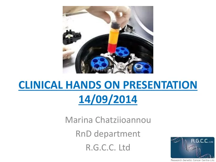

CLINICAL HANDS ON PRESENTATION 14/09/2014 Marina Chatziioannou RnD department R.G.C.C. Ltd
Points of interest 1. CTCs isolation − Manual or automated cell isolation − Magnetic bead epitope selection 2. Clinical flow cytometry − What for? − Epitope selection − Sample handling − Data analysis
CTCs isolation Topics 1. Sample arrival and handling 2. Manually or automated cell isolation 3. Magnetic bead epitope selection 4. Culture treatment
CTCs isolation Manually Ficoll layering Washing Beads incubation Magnetic CTCs isolation Culture in FL25 Automated Robosep handling Culture in FL25
Epitopes (bead selection) Categorization 1. Epithelial origin 2. Hematological origin 3. Sarcomas 4. Non epithelial, non hematological, non sarcoma
Epithelial origin EpCAM i. Expressed exclusively in epithelia and epithelial-derived neoplasms. ii. Can be used as diagnostic marker for various cancers. iii. Plays a role in tumorigenesis and metastasis of carcinomas, so it can also act as a potential prognostic marker.
Hematological origin CD45 i. Leukocyte Common Antigen (LCA). ii. Its various forms are present on all differentiated hematopoietic cells except erythrocytes and plasma cells. iii. It is expressed in lymphomas, B-cell chronic lymphocytic leukemia, hairy cell leukemia, and acute nonlymphocytic leukemia. iv. A monoclonal antibody to CD45 is used in routine immunohistochemistry to differentiate between histological sections from lymphomas and carcinomas.
Sarcomas CD99 i. Ewing’s marker ii. Present to all sarcomas
Non epithelial, non hematological, non sarcomas CD45 negative selection i. For types of cancer that are not included in the previous categorizations. ii. For example: • Renal cancer • SCLC • NK lymphomas • Neuroblastomas • Adrenal cancer
Flow cytometry for clinical samples Topics • What for? • Epitopes selected • Sample handling • Data analysis
What for? • Easy, quick and reliable technique. • Gives accurate results. • Quick view of what to expect in the isolated culture from the same patient.
Flow cytometry clinical epitopes i. CD45:LCA ii. CD31: Endothelial cell marker iii. cMet: Metastatic cell marker iv. PanCK: Marker of cell of origin of various tumors (depends on cytokeratis pattern) v. CD227: Breast, Lung cancer marker vi. CD63: Melanoma cancer cell marker vii. PSMA: Prostate cancer cell marker
Sample handling Steps 1. Extracellular staining 2. Washing 3. Fixation 4. Washing 5. Permeabilization 6. Intracellular staining
Data analysis • Flow cytometry data file is analyzed and percentages of epitopes are produced. • Results are represented as below:
References 1. Ignatiadis, M., C. Sotiriou, et al. (2012). "Minimal residual disease and circulating tumor cells in breast cancer: open questions for research." Recent Results Cancer Res 195 : 3-9. 2. Gendler SJ (July 2001). "MUC1, the renaissance molecule". J. Mammary Gland Biol Neoplasia 6 (3): 339 – 353. doi:10.1023/A:1011379725811. PMID 11547902 3. Radford KJ, Thorne RF, Hersey P (1996). "CD63 associates with transmembrane 4 superfamily members, CD9 and CD81, and with beta 1 integrins in human melanoma". Biochem. Biophys. Res. Commun. 222 (1): 13 – 18. doi:10.1006/bbrc.1996.0690. PMID 8630057 4. Omary MB, Ku NO, Strnad P, Hanada S (July 2009). "Toward unraveling the complexity of simple epithelial keratins in human disease". J. Clin. Invest. 119 (7): 1794 – 805. doi:10.1172/JCI37762. PMC 2701867. PMID 19587454 5. Tang DG, Chen YQ, Newman PJ, et al. (1993). "Identification of PECAM-1 in solid tumor cells and its potential involvement in tumor cell adhesion to endothelium.". J. Biol. Chem. 268 (30): 22883 – 94. PMID 8226797 6. Gentile A, Trusolino L, Comoglio PM (March 2008). "The Met tyrosine kinase receptor in development and cancer". Cancer Metastasis Rev. 27 (1): 85 – 94. doi:10.1007/s10555-007- 9107-6. PMID 18175071
Thank you for your time and patience!
Recommend
More recommend