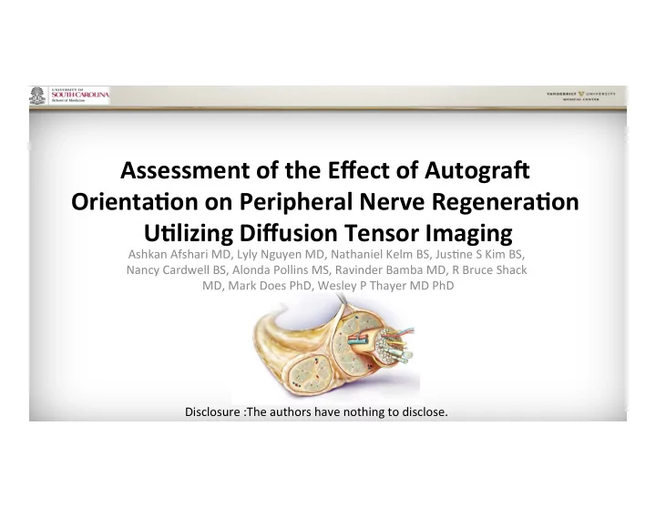

Assessment ¡of ¡the ¡Effect ¡of ¡Autogra2 ¡ Orienta5on ¡on ¡Peripheral ¡Nerve ¡Regenera5on ¡ U5lizing ¡Diffusion ¡Tensor ¡Imaging ¡ Ashkan ¡Afshari ¡MD, ¡Lyly ¡Nguyen ¡MD, ¡Nathaniel ¡Kelm ¡BS, ¡Jus;ne ¡S ¡Kim ¡BS, ¡ Nancy ¡Cardwell ¡BS, ¡Alonda ¡Pollins ¡MS, ¡Ravinder ¡Bamba ¡MD, ¡R ¡Bruce ¡Shack ¡ MD, ¡Mark ¡Does ¡PhD, ¡Wesley ¡P ¡Thayer ¡MD ¡PhD ¡ Disclosure ¡:The ¡authors ¡have ¡nothing ¡to ¡disclose. ¡
Nerve ¡AutograI ¡ • Few ¡studies ¡to ¡evaluate ¡the ¡effect ¡of ¡autograI ¡ polarity. ¡ – Limited ¡by ¡number ¡of ¡assessment ¡tools. ¡ • No ¡consensus ¡on ¡role ¡of ¡autograI ¡orienta;on. ¡ Stromberg B V, Vlastou C, Earle A S. Effect of nerve graft polarity on nerve regeneration and function. J Hand Surg . 1979;4:444-5. Sotereanos DG, Seaber AV, Urbaniak JR. Reversing nerve-graft polarity in a rat model: the effect on function. J Reconstr Microsurg . 1992;8:303-7. Nakatsuka H, Takamatsu K, Koshimune M, et al. Experimental study of polarity in reversing cable nerve grafts. J Recontr Microsurg . 2002;18(6):509-15. Ansselin AD, Davey DF. The regeneration of axons through normal and reversed peripheral nerve grafts. Restor Neurol Neurosci . 1993;5(3):225-40.
Purpose ¡ • Evaluate ¡the ¡effect ¡of ¡autograI ¡orienta;on ¡on ¡ nerve ¡recovery ¡using ¡mul;ple ¡assessments ¡ tools, ¡including ¡DTI. ¡
Methods ¡(Design) ¡ 36 ¡Sprague-‑Dawley ¡ female ¡rats ¡ 12 ¡Reverse ¡ 12 ¡Normal ¡ 12 ¡Sham ¡ Orienta;on ¡ Orienta;on ¡ Weekly behaviors for 6 weeks Gastrocnemius/Soleus ¡Muscle ¡Harvest ¡ Le2 ¡Scia5c ¡Nerve ¡Harvest ¡ DTI ¡ IHC/TB ¡
Methods ¡(Microsurgery) ¡ Sham Normal Orientation Reverse Orientation 10mm 10mm p d
Behavior: ¡Scia;c ¡Func;on ¡Index ¡ ¡ ¡ • Hind ¡limbs ¡inked ¡and ¡animal ¡ walks ¡up ¡an ¡inclined ¡plank ¡ ¡ • Markings ¡measured ¡and ¡ inserted ¡into ¡a ¡validated ¡ formula ¡ • Greater ¡impairment ¡ demonstrated ¡by ¡more ¡ nega;ve ¡score ¡
Behavior: ¡Foot ¡Fault ¡ • Animals ¡allowed ¡to ¡take ¡50 ¡steps/hind ¡limb ¡on ¡wired ¡ grid, ¡and ¡number ¡of ¡foot ¡faults ¡(FF) ¡recorded ¡ – Par;al ¡FF ¡(through ¡grid ¡without ¡touching ¡base) ¡= ¡1 ¡point ¡ – Full ¡FF ¡(through ¡grid ¡and ¡touches ¡base)= ¡2 ¡points ¡ Foot ¡Fault ¡Asymmetry ¡Score = ¡%foot ¡ • fault ¡(surgical ¡hind ¡limb) ¡-‑ ¡%foot ¡fault ¡ (normal ¡hind ¡limb) ¡ 2cm
Muscle ¡Net ¡Weight ¡ • Net ¡weight ¡(gm)= ¡weight ¡(normal ¡limb ¡gastrocnemius/soleus ¡ m.)-‑ ¡weight ¡(surgical ¡limb ¡gastrocnemius/soleus ¡m.) ¡
Histology ¡ Immunohistochemistry ¡ Toluidine ¡Blue ¡ • 5 ¡μm ¡thick, ¡Choline-‑ • 1 ¡μm ¡thick ¡sec;ons ¡ acetyltransferase ¡(ChAT) ¡ • Axon ¡count, ¡density ¡ stained ¡for ¡motor ¡axon ¡ and ¡diameter ¡at ¡40X ¡ counts ¡at ¡10X ¡
Diffusion ¡Tensor ¡Imaging ¡(DTI) ¡ • Common ¡tool ¡used ¡in ¡ evalua;on ¡of ¡CNS; ¡emerging ¡ MRI ¡technique ¡for ¡PNS. ¡ • Relies ¡on ¡diffusion ¡of ¡water ¡ molecules ¡within ¡;ssue. ¡ • Frac;onal ¡anisotropy, ¡axial ¡ and ¡radial ¡diffusivity, ¡and ¡ tractography ¡data ¡obtained. ¡ Lehmann HC, Zhang J, Mori S, Sheikh KA. Diffusion tensor imaging to assess axonal regeneration in peripheral nerves. Exp Neurol . 2010;223(1):238-44. Sheikh KA. Non-invasive imaging of nerve regeneration. Exp Neurol . 2010;223(1):72-6.
Results: ¡Behavior ¡studies ¡ Foot Fault SFI 5 ¡ 20 ¡ 0 ¡ Scia5c ¡Func5on ¡Index ¡Score ¡ 0 ¡ -‑5 ¡ Foot ¡Fault ¡Score ¡ -‑20 ¡ -‑10 ¡ -‑40 ¡ -‑15 ¡ Sham ¡ -‑60 ¡ -‑20 ¡ Normal ¡Orienta;on ¡ -‑80 ¡ -‑25 ¡ Reverse ¡Orienta;on ¡ -‑100 ¡ -‑30 ¡ -‑120 ¡ -‑35 ¡ 72 ¡ 1 ¡week ¡ 2 ¡ 3 ¡ 4 ¡ 5 ¡ 6 ¡ 72 ¡ 1 ¡week ¡ 2 ¡ 3 ¡ 4 ¡ 5 ¡ 6 ¡ hours ¡ weeks ¡ weeks ¡ weeks ¡ weeks ¡ weeks ¡ hours ¡ weeks ¡ weeks ¡ weeks ¡ weeks ¡ weeks ¡ Postopera5ve ¡Time ¡ Postopera5ve ¡Time ¡ *No ¡difference ¡in ¡FF ¡or ¡SFI ¡between ¡normal ¡and ¡reverse ¡autogra9s ¡ ¡
Results: ¡Muscle ¡Net ¡Weight ¡ 1.6 ¡ 1.36 ¡ 1.33 ¡ 1.4 ¡ 1.2 ¡ Weight ¡(gm) ¡ 1 ¡ Sham ¡ 0.8 ¡ Normal ¡Orienta;on ¡ 0.6 ¡ Reverse ¡Orienta;on ¡ 0.4 ¡ 0.2 ¡ 0.02 ¡ 0 ¡ *No ¡difference ¡between ¡ Sham ¡ Normal ¡Orienta5on ¡ Reverse ¡Orienta5on ¡ ¡autogra9 ¡groups ¡
Results: ¡Motor ¡IHC ¡ Proximal Graft Distal 1600 ¡ 1400 ¡ Motor ¡Axon ¡Count ¡ 1200 ¡ 1000 ¡ Sham Sham ¡ 800 ¡ 600 ¡ Normal ¡ Orienta;on ¡ 400 ¡ Reverse ¡ 200 ¡ Normal Orienta;on ¡ 0 ¡ Orientation Proximal ¡ Gra2 ¡ Distal ¡ Nerve ¡Segment ¡ *No ¡difference ¡in ¡motor ¡axon ¡count ¡between ¡ Reverse normal ¡and ¡reverse ¡autogra9s ¡at ¡any ¡nerve ¡segment ¡ ¡ Orientation
Results: ¡Toluidine ¡Blue ¡ 8630 ¡ 8429 ¡ Graft Distal 10000 ¡ 6778 ¡ 7547 ¡ 7668 ¡ Axon ¡Counts ¡ 6778 ¡ 5000 ¡ 0 ¡ Sham 10000 ¡ 6655 ¡ 7739 ¡ 7429 ¡ Axon ¡Density ¡ (axons/mm 2 ) ¡ 6655 ¡ 5812 ¡ 5905 ¡ 8000 ¡ 6000 ¡ 4000 ¡ Normal 2000 ¡ Orientation 0 ¡ 8.74 ¡ 8.74 ¡ 10 ¡ Axon ¡Diameter ¡ Reverse 5.16 ¡ 5.09 ¡ 3.51 ¡ 3.92 ¡ 5 ¡ Orientation (μm) ¡ 0 ¡ *No ¡difference ¡in ¡axon ¡count, ¡density ¡or ¡diameter ¡between ¡normal ¡and ¡reverse ¡ autogra9s ¡within ¡and ¡distal ¡to ¡the ¡autogra9 ¡
Results: ¡DTI ¡ Comparison ¡of ¡DTI ¡parameters ¡between ¡normal ¡and ¡reverse ¡autogra2s ¡at ¡all ¡nerve ¡segments ¡ Sham Reverse Normal Proximo-‑distal ¡axonal ¡growth ¡demonstrated ¡ ¡in ¡normal ¡and ¡reverse ¡autogra9s ¡ *No ¡difference ¡in ¡FA, ¡AD, ¡RD ¡between ¡normal ¡and ¡reverse ¡autogra9s ¡ ¡at ¡any ¡nerve ¡segment ¡
Conclusion ¡ • Nerve ¡regenera;on ¡was ¡similar ¡in ¡reverse-‑ ¡and ¡ normal-‑oriented ¡autograIs. ¡ • AutograI ¡polarity ¡may ¡not ¡influence ¡nerve ¡ regenera;ve ¡outcomes. ¡ • Nerve ¡repairs ¡u;lizing ¡non-‑branched ¡autograIs ¡ should ¡be ¡performed ¡using ¡principles ¡(i.e. ¡best ¡ fascicular ¡alignment) ¡other ¡than ¡orienta;on ¡to ¡ maximize ¡regenera;on. ¡ ¡
Recommend
More recommend