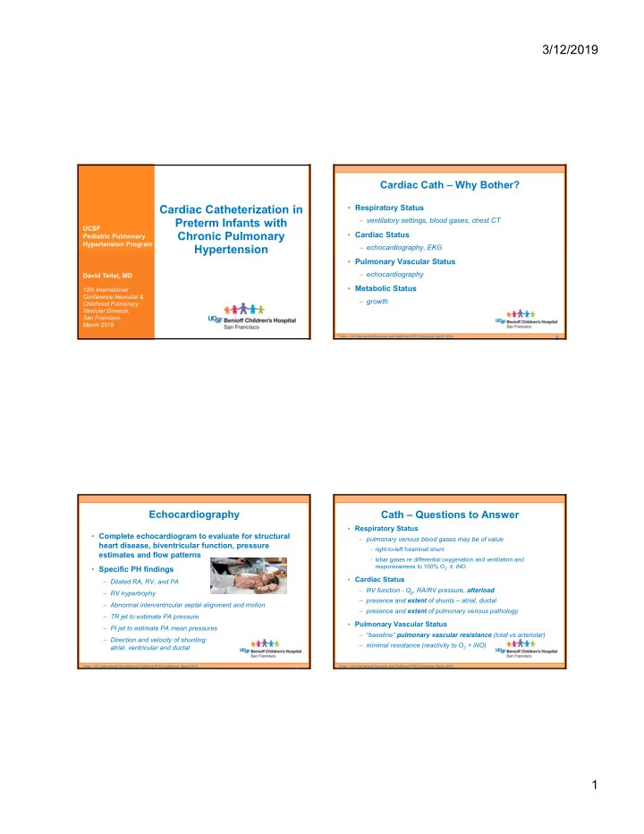

3/12/2019 Cardiac Cath – Why Bother? Cardiac Catheterization in • Respiratory Status Preterm Infants with – ventilatory settings, blood gases, chest CT UCSF Chronic Pulmonary • Cardiac Status Pediatric Pulmonary Hypertension Program Hypertension – echocardiography, EKG • Pulmonary Vascular Status – echocardiography David Teitel, MD • Metabolic Status 12th International Conference Neonatal & – growth Childhood Pulmonary Vascular Disease, San Francisco, March 2019 Teitel – 12 th International Neonatal and Childhood PVD Conference, March 2019 2 Echocardiography Cath – Questions to Answer • Respiratory Status • Complete echocardiogram to evaluate for structural – pulmonary venous blood gases may be of value heart disease, biventricular function, pressure • right-to-left foraminal shunt estimates and flow patterns • lobar gases re differential oxygenation and ventilation and responsiveness to 100% O 2 ± iNO • Specific PH findings • Cardiac Status – Dilated RA, RV, and PA – RV function - Q p , RA/RV pressure, afterload – RV hypertrophy – presence and extent of shunts – atrial, ductal – Abnormal interventricular septal alignment and motion – presence and extent of pulmonary venous pathology – TR jet to estimate PA pressure • Pulmonary Vascular Status – PI jet to estimate PA mean pressures – “baseline” pulmonary vascular resistance (total vs arteriolar) – Direction and velocity of shunting: – minimal resistance (reactivity to O 2 + iNO) atrial, ventricular and ductal Teitel – 12 th International Neonatal and Childhood PVD Conference, March 2019 Teitel – 12 th International Neonatal and Childhood PVD Conference, March 2019 3 1
3/12/2019 Cath – Problems to Fix Cath – Considerations & Caveats • “Baseline state” • During cath – effect of anesthesia, necessity to increase ventilatory support – device closure of PDA • Qp (Qs often equals Qp and less important to questions) – device closure of ASD – if Fick, need to assume oxygen consumption – pulmonary vein dilation • thermodilution when possible, and back-calculate VO 2 – mixed pulmonary venous saturation in presence of atrial r-l shunt • Post cath – mixed pulmonary arterial saturation with l-r ductal shunt – minimize RV afterload with a goal to reach minimal cath PVR – importance of including dissolved oxygen when pO2 ≥ 150 mmHg with therapy to assist in vascular remodeling • Pulmonary vascular resistance – what is normal for a premature infant? – how does one correct for damaged lung parenchyma? Teitel – 12 th International Neonatal and Childhood PVD Conference, March 2019 Teitel – 12 th International Neonatal and Childhood PVD Conference, March 2019 Oxygen Consumption Oxygen Consumption Seckeler MD, et al. A new predictive equation for oxygen consumption in children and adults Seckeler MD, et al. A new predictive equation for oxygen consumption in children and adults with congenital and acquired heart disease. Heart. 2015;101(7):517-24. with congenital and acquired heart disease. Heart. 2015;101(7):517-24. Teitel – 12 th International Neonatal and Childhood PVD Conference, March 2019 Teitel – 12 th International Neonatal and Childhood PVD Conference, March 2019 2
3/12/2019 Patient 1 Patient 2 • 7 mo, former 23 wk premie, • 7 mo, former 23 wk premie, • 10 mo, former 25 wk premie,multiple • 10 mo, former 25 wk premie,multiple transferred from OSH with multiple transferred from OSH with multiple complications, transferred from OSH complications, transferred from OSH complications, tracheostomy on complications, tracheostomy on on high flow O 2 , on sildenafil on high flow O 2 , on sildenafil ventilator, on sildenafil ventilator, on sildenafil • Echo: RVP = ½ systemic, RVH, • Echo: RVP = ½ systemic, RVH, • Echo: RVP = ½ systemic, no RVH, • Echo: RVP = ½ systemic, no RVH, small PDA, no atrial communication small PDA, no atrial communication no TR, atrial septum not well seen no TR, atrial septum not well seen • 19 mo, off oxygen, no vasodilators, • 23 mo, still trach/vent, on triple g-tube feeds therapy with sc Remodulin • Echo: none since 2 months post • Echo: normal RVP, no RVH, no cath – still mild RVH atrial communication (normal echo) Teitel – 12 th International Neonatal and Childhood PVD Conference, March 2019 9 Teitel – 12 th International Neonatal and Childhood PVD Conference, March 2019 10 Patient 3 Summary • Diagnostic value of cath • 10 wo, former 30 wk premie, • 10 wo, former 30 wk premie, transferred from OSH with multiple transferred from OSH with multiple – pulmonary vascular resistance, “baseline” and minimal , can be anomalies, CLD on high flow O 2 anomalies, CLD on high flow O 2 accurately measured, and cannot be accurately estimated from echocardiography, even when pressure can be estimated • Echo: RVP > ½ systemic, RVH, • Echo: RVP > ½ systemic, RVH, PDA and atrial level shunt PDA and atrial level shunt • corollary: can differentiate the impact of increases in pulmonary blood flow versus vascular resistance on RV pressure estimated by echo • 6 mo, hospitalized, on bosentan & – can independently evaluate individual pulmonary venous anatomy sildenafil, high flow O 2 , g-tube and lobar pulmonary function (oxygenation and ventilation) • Echo: RVP > ½ systemic, RVH, • Therapeutic value of cath mild RA & RV dilation, fenestrated – can intervene on shunts when they are the source of elevated RVP atrial septum – can intervene on pulmonary veins (if you are so pre-disposed) Teitel – 12 th International Neonatal and Childhood PVD Conference, March 2019 Teitel – 12 th International Neonatal and Childhood PVD Conference, March 2019 11 3
Recommend
More recommend