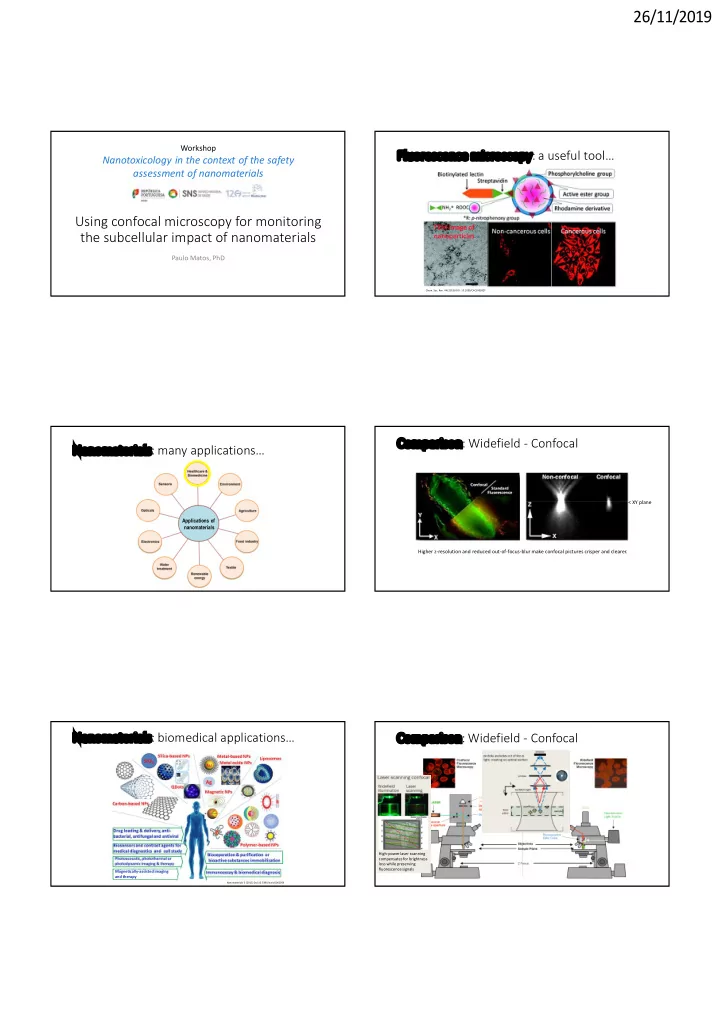

26/11/2019 Workshop Fluorescence microscopy: a useful tool… Nanotoxicology in the context of the safety assessment of nanomaterials Using confocal microscopy for monitoring the subcellular impact of nanomaterials Paulo Matos, PhD Chem. Soc. Rev. 44 (2015) DOI: 10.1039/C4CS00392F Comparison: Widefield - Confocal Nanomaterials: many applications… < XY plane Applications of nanomaterials Higher z-resolution and reduced out-of-focus-blur make confocal pictures crisper and clearer. Nanomaterials: biomedical applications… Comparison: Widefield - Confocal High-power laser scanning compensates for brightness loss while preserving fluorescence signals Magnetically-assisted imaging and therapy Nanomaterials 5 (2015) Doi:10.3390/nano5042054
26/11/2019 Comparison: Widefield - Confocal Confocal: Z-sectioning Increased Z-scanning Stack of XY images (Z-sections) 3D-reconstruction A= XY middle section B, C = XZ side views at different Y-positons Z < Y Y Z X Y Confocal: Localization of nanomaterials in cells Widefield: optical section NP Widefield fluorescence microscopy NP (ZX) Nanoparticles (NP) EEA1 The precise location of a signal relatively to the 3D volume of the cell cannot be accurately Confocal determined! (XY) LAMP1 TEM Signal CRC cells exposed to cellulose nanofibers Confocal: optical section (ZX) There is no superimposition of out-of-focus (Z-plane) signals! The location of a Propidium iodide (PI) signal relatively to the Calcofluor white (CW) 3D volume of the cell can be accurately determined! (XY)
26/11/2019 CRC cells exposed to cellulose nanofibers Polarized Caco2:HT29-Mtx co-culture exposed to titanium dioxide nanomaterials (TiO 2 ) Apical surface plane XY XY Actin DAPI TiO 2 NM Propidium iodide (PI) Calcofluor white (CW) YZ XZ CRC cells exposed to cellulose nanofibers Time course monitoring of live HeLa cells exposed to 2 different NMs Cell membranes stained with CellMask DeepRed Larger NPs exert interactions with the cell membrane that are sufficiently strong to trigger D-penicillamine-coated quantum dots (DPA-QDs) rapid internalization…. Z-scanning Dihydrolipoic acid-coated gold nanoclusters (DHLA-AuNCs) Smaller NPs have to form a cluster of a certain size to induce membrane invagination, delaying internalization… Beilstein J. Nanotechnol. 5 (2014) DOI:10.3762/bjnano.5.248 CRC cells exposed to cellulose nanofibers What is the fate of internalized NPs? NP Early endosomes EEA1 EEA1 DAPI Rab14 LAMP1
26/11/2019 Following macrophages’ endocytic trafficking of crystalline What is the fate of internalized NPs? antiretroviral nanoparticles (RTV-NPs)… NP Late endosomes EEA1 Rab7 DAPI Rab14 LAMP1 Gendelman et al. NANOMEDICINE 6 (2011) DOI: 10.2217/nnm.11.27 What is the fate of internalized NPs? Lysosomal entrapment of NMs…a road block? NP Delivery Lysosomes to specific intracellular Compartment? EEA1 LAMP1 Rab14 LAMP1 Most common route for internalized NMs, but… Target cancer cells What is the fate of internalized NPs? How to monitor NMs’ lysosomal escape? NP NMs can be developed or optimized to Recycling endosomes promote of endosomal escape : (a) Membrane fusion between the nanoparticle structure and the endosomal EEA1 membrane to release the cargo into the cytosol. Rab11 (b) The proton sponge mechanism where the NMs’ buffering of lysosome pH increases the ionic influx of chloride osmotic lysing the lysosome Rab14 (c) Swelling of pH responsive nanogels ruptures the lysosomal membrane through mechanical strain. LAMP1 (d) pH responsive nanoparticles disassemble and destabilize the lysosomal membrane. BIOCONJUGATE CHEM 30 (2019) DOI: 10.1021/acs.bioconjchem.8b00732
26/11/2019 Confocal microscopy to monitor NMs’ Co-localization with organelle-specific markers… lysosomal escape… Mitochondria Dyes that change fluorescence intensity in F 1 - ATPase response to a shift in pH DAPI (e.g., fluorescein, calcein): can be coupled to NPs to monitor their escape from acidic lysosomes to the cytosol (pH ∼ 7.2) by fluorescence microscopy. BIOCONJUGATE CHEM 30 (2019) DOI: 10.1021/acs.bioconjchem.8b00732 Confocal microscopy to monitor NMs’ Co-localization with organelle-specific markers… lysosomal escape… Golgi-apparatus GM130 DAPI GFP complementation assay : A small fragment of GFP is conjugated through a linker to a material. Fluorescence only appears if If the material escapes and complements with the large GFP fragment in the cytosol. Co-localization with organelle-specific markers… Co-localization with organelle-specific markers… Endoplasmic Reticulum Calnexin DAPI The Golgi-targeting ability of L-cysteine LC-F-SiO 2 GM130 functionalized silica nanoparticles. Chemical Science 8 (2017) DOI:10.1039/C7SC01316G
26/11/2019 Visualizing NMs’ interaction with the Acknowledgements cytoskeleton… Microtubules Toxicology Lab Oncobiology Lab Maria João Silva Peter Jordan Henriqueta Louro Joana Pereira Célia Ventura Tubulin Funding Fátima Pinto DAPI Dora Rolo Ana Gramacho Sara Teixeira Carbon nanotubes cause DNA bridges in LC cells Merge Tubulin DAPI Z XY plane Ventura et al., in press Manipulating microtubules to relax the endothelial barrier… Using confocalmicroscopy for monitoring the subcellular impact of nanomaterials Paulo Matos, INSARJ This makes the endothelial barrier “leaky” at specific sites, to allow drug molecules to pass The physicochemical properties of nanomaterials, such as their small size and high surface area ratio, make Fluorescent iron oxide nanoparticles into tissues… them ideal for many applications in industry and biomedicine. However, those same properties increase their ability to interact with cells and tissues, allowing their permeation through several biological barriers. While bind microtubules in endothelial cells... these abilities have been exploited in the development of novel drug-delivery systems, the widespread use of nanomaterials makes the evaluation of the potential cytotoxicity of their raw materials an important public health issue. In vivo studies are the usual gold standard when assessing compound toxicity, however, in vitro studies have also provided a lot of information regarding the toxicity and MoA of many compounds, and have proved crucial to clarify how the intrinsic and extrinsic properties of certain nanomaterials contribute to their interaction with cells an tissues. In this talk we will describe how confocal microscopy can be used in in vitro cell cultures to evaluate the subcellular impact of nanomaterials. We will point out the advantages and limitations of using confocal fluorescent microscopy in investigating how cells interact and react to the presence of different types of nanomaterial and how these can affect basic cellular functions. Under a magnetic field (MF), the nanoparticles re-align the microtubule structure, temporarily disrupting cell–cell junctions… Appl Nanosci 4 (2014) DOI: 10.1007/s13204-013-0216-y https://www.genengnews.com/topics/drug-discovery/leaks-magnetically-induced-between-cells-could-promote-drug-delivery/
Recommend
More recommend