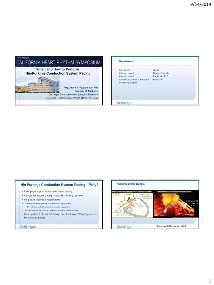

9/14/2019 Disclosures When and How to Perform Consultant - Abbott His-Purkinje Conduction System Pacing Advisory board - Boston Scientific Advisory board - Eaglepoint LLC Speaker, Consultant, Research - Medtronic Fellowship support Pugazhendhi Vijayaraman, MD Professor Of Medicine Geisinger Commonwealth School of Medicine Geisinger Heart Institute, Wilkes-Barre, PA, USA 1 2 His Purkinje Conduction System Pacing – Why? Anatomy of His Bundle • Most physiological form of ventricular pacing • Conduction occurs through native His-Purkinje system • No pacing induced dyssynchrony LV-RV part of MS • Lead tip and body potentially within the right atrium LV-RA part of MS – Prevents lead related issues such as tricuspid regurgitation HB • Both AV and VV synchrony can be achieved at the same time • Has significant clinical advantage over traditional RV pacing or even AV node biventricular pacing Courtesy: K.Shivkumar, UCLA 3 1
9/14/2019 Selective His Bundle Pacing Histological 90 90 40 40 S V H V HBP Correa de Sa,…, Lustgarten D. Circ AE 2012 5 6 Nonselective HBP Nonselective HBP Baseline HBP 1.5V 1.0V Baseline HBP 1.2V 1.0V 80 120 160 90 140 90 A A 40 250 150 RA 40 HBP H H V 150 150 V RA 50 HBP H 2
9/14/2019 ECG IMAGING Patient Selection for HPCSP • AV nodal block • Infra-nodal AV block • Sinus node dysfunction • Atrial fibrillation with slow ventricular rate • AV node ablation • Cardiac Resynchronization Therapy Vijayaraman P, et al. His bundle pacing JACC 2018;72:927-47 9 10 Overview of Implantation Overview of Implantation • • Deflectable delivery sheath Fixed Curve Medtronic C315 His sheath • Medtronic SelectSite C304 - 8 Fr 11 12 3
9/14/2019 • Deflectable Medtronic C304His sheath C315His C304His C304 13 14 Overview of Implantation Overview of Implantation • Electrophysiology mapping catheter to locate the His A A V H H Unipolar mapping 15 16 4
9/14/2019 How to perform HBP 0.1 mV/mm HBP 0.5V @ 0.5ms 18 19 20 5
9/14/2019 His bundle injury current His bundle injury current At implant IC No IC P value Number of pts 60 (n, %) 22 (37%) 22 (37%) 38 (63%) 38 (63%) Fluoroscopy duration ( min) 8.9 ± 4 9.5 ± 3.5 NS R wave (mV) 4.1 ± 2.8 5.4 ± 3.2 NS 5 minutes Pacing Impedance (Ohms) 557 ± 97 639 ± 159 NS Pacing thresholds (V @ 0.5 ms) Mean ± SD Mean ± SD Implant 1.16±0.4 1.16±0.4 1.75±0.7 1.75±0.7 <0.05 2 weeks 1.18±0.5 1.18±0.5 1.82±0.8 1.82±0.8 <0.05 2 months 1.23±0.6 1.23±0.6 1.93±0.8 1.93±0.8 <0.05 20 minutes 1 yr (N =41) 1.31±0.6 1.31±0.6 1.98±0.9 1.98±0.9 <0.05 Vijayaraman P,Dandamudi G, Worsnick SA, et al. Acute His bundle injury…. PACE 2015:38:540-6 Complete AV nodal block 2:1 HV block A A A A H H H H H V V H H A A A A A A 6
9/14/2019 Complete AV Block Mapping the His bundle in the setting of Intra-Hisian Block | 25 Proximal His Distal His H H H H H H A A A A 7
9/14/2019 His bundle Mapping Output Dependent His Capture 1.4V @ 1 ms 1.2V @ 1 ms 1.0V @ 1 ms Proximal His Bundle Distal His Bundle NS-HBP S-HBP S-LBP His Bundle Pacing in Advanced AV block Advanced AV block N = 100 AV nodal Block Infra nodal Block N = 46 N = 54 HBP and AV Node ablation Successful Unsuccessful Successful Unsuccessful 43 (93%) 3 (7%) 41 (76%) 13 (24%) Vijayaraman P, Naperkowski A, Ellenbogen KA et al. JACCEP 2015;1:571-81 31 32 8
9/14/2019 AV Node Ablation Site in Relation to HBP HBP and AVNA Electrodes • HBP and AVNA were performed simultaneously in 30 patients (71%) AORTA • AVNA in patients with prior HBP in 8 (19% - 1 to 15 months after) * * * T (near Tip) • HBP in patients with previous AVNA in 4 (10% - infection 2, HF 2; 1-12 * TR (Tip to Ring) * HBP lead * * * * years after) * * R (at Ring) * * * * * * * * * * * TV * BR (Below Ring) * * * * * * * * • * * L (Left sided) HBP and AVNA was successful in 40/42 patients (95%) * * * * * * * * * • HBP lead placement was unsuccessful in one pt. • HBP lead dislodged during failed attempt at AVN ablation on the right side → left sided ablation CS Os Vijayaraman P et al. Europace 2017;19:iv10-16 33 Vijayaraman P et al. Europace 2017;19:iv10-16 Left ventricular Ejection Fraction (%) P = 0.5 P = 0.01 60 P < 0.001 50 40 Baseline Post-HBP 30 20 HBP for Cardiac Resynchronization Therapy 10 0 LVEF (all) LVEF <40% LVEF >40% 35 36 Vijayaraman P, Subzposh F, Naperkowski A. Europace 2017;19:iv10-16 9
9/14/2019 Heart Rhythm 2018;15:413-420 LBBB Selective His Bundle Pacing @ 2 V Selective HBP @ 1.4 V C B C 190 95 190 AV node AV node AV node HBP HBP HBP lead lead lead 37 38 95/106 (90%) 35 Methods 31 Group 1 Group 2 30 Permanent HBP was attempted in patients with cardiomyopathy, 27 reduced LV function (EF <50%) and heart failure 25 25 25 Attempted 25 Cases 22 Group I (RESCUE HBP) 20 • LV lead placement was unsuccessful 15 15 • Prior CRT did not result in clinical response (non-responder) 15 Successful Cases Group II (PRIMARY HBP) 10 8 8 • AV block / AV node ablation 5 • High RV pacing burden • Bundle branch block 0 Rescue HBP in Rescue HBP in BiV Primary HBP in Primary HBP in Primary HBP in 39 Failed BiV Non-responders AVB/AVJ BBB Ventricular Pacing 40 10
9/14/2019 QRS duration LV Ejection Fraction * P < 0.001 Baseline QRSd HBP QRSd Baseline Follow-up 200 * p-value = 0.0001 60 177 55 180 163 157 50 160 * 44 44 * 140 40 125 40 118 p = 0.04 116 120 108 103 30 30 100 25 80 20 60 40 10 20 0 0 Overall Baseline LVEF < 35% Baseline LVEF 35-50% Overall BBB Ventricular paced Narrow QRS Ahran D. Arnold et al. JACC 2018;72:3112-3122 41 HBP IN RBBB Sharma PS, Naperkowski A, Bauch T, Chan DSY , Whinnett Z, Arnold A, Ellenbogen KA, Vijayaraman P. | 44 11
9/14/2019 Baseline RBBB Nonselective HBP Selective HBP Selective RBP ECG Imaging Figure 2 5 V @ 1 ms 1.5 V @ 1 ms 1.0 V @ 1 ms RV Selective-RBP His bundle Nonselective-HBP Selective-HBP Intra-Hisian RBBB HBP lead HBP lead HBP lead QRS duration NYHA Class Ejection Fraction 158 Vijayaraman P, Herweg B, Ellenbogen KA, Gajek J. 2019;12:e006934 * 39 160 3.5 40 150 2.8 * 3 140 35 127 31 130 2.5 * 2.0 120 30 2 110 100 25 1.5 90 80 20 1 Baseline HBP Baseline HBP Baseline HBP * P <0.001 47 48 12
9/14/2019 83 yrs old man with ischemic CMP, LBBB, Class IV CHF Selective HBP His Optimized CRT His synchronus LV pacing (His-LV timing at 50 ms) 95% echocardiographic response His → LV 50 ms Baseline S-HBP QRS Duration P < 0.001 200 * vs baseline * vs BVP 180 210 146 110 * * vs HBP ** 146 160 210 110 140 *** 120 100 LV 80 Q-LV 160 ms 96 ms 0 Baseline BVP HBP HOT-CRT QRSd 210 ms 146 ms 110 ms I LEFT BUNDLE BRANCH AREA PACING LBB pacing can be easily achieved? II III Narrow target aVR HB accurate positioning needed aVL aVF Wider conduction LB net V1 Right bundle B Easy to find and fix V2 His bundle V3 RB V4 B V5 V6 LB AV node LBBP H Left bundle HBP 52 Vijayaraman P et al. Heart Rhythm 2019 13
9/14/2019 Case presentation AV nodal HB • 75-year-old man LBB • Prior CAD, s/p PCI • Chronic LBBB x 20 years • LVEF 40% on OMT x 20 years • Syncope → intermittent CHB 53 54 54 Baseline NSHBP LBBP-Uni Bipolar 3V 0.6V 0.5V I II III aVR aVL aVF V1 V2 V3 V4 V5 V6 LBBP H HBP 14
9/14/2019 Baseline AVD 200 ms 170 ms 150 ms 130 ms 100 ms 80 ms I LBBP lead II III aVR aVL aVF contrast HBP V1 V2 LV LBB V3 P V4 V5 V6 LBBP HBP RA BBB/IVCD AV block His bundle pacing No correction or Complete BBB Incomplete BBB no His capture correction correction Consider addition Standard BIV His lead only of LV lead yes BBB correction pacing / CRT HOT CRT at <2V Left Bundle ?Left Bundle branch branch Pacing Pacing 59 15
9/14/2019 Conclusions ➢ Permanent HPCSP is feasible in all patients requiring permanent pacemakers. Thank you for your attention ➢ HPCSP is effective in patients requiring AV node ablation ➢ HPCSP may be considered as an alternative to biventricular pacing in patients requiring CRT #DontDisTheHis 61 62 16
Recommend
More recommend