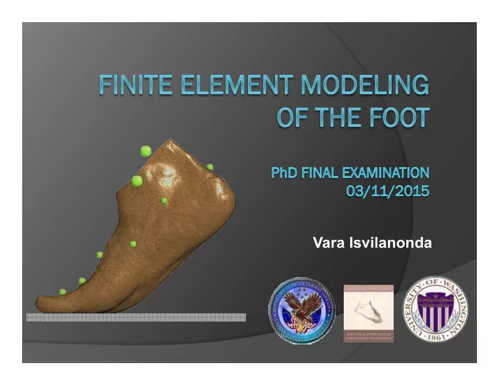

Vara Isvilanonda
Specific Aim 4: Introduction Objective To develop and validate two subject-specific FE foot models (normal and diabetic), to explore the plantar pressure and internal soft tissue stress during quiet stance and the stance phase of gait, and to investigate the effect of soft tissue assumptions.
Specific Aim 4: Introduction Normal and diabetic subjects ! Normal subject " Age 43 year-old (male) " Body weight 945 N ! Diabetic subject " Age 31 year-old (male) " Body weight 688 N " Duration of diabetes > 25 years
Specific Aim 4: Method FE foot model development Obtain imaging data CT (10% BW per foot) MRI (unloaded) Bone anatomy Skin, fat and muscle anatomy
Specific Aim 4: Method Segmentation S L M L I S P A I A P
Specific Aim 4: Method Boolean operations Generate joint cavities Generate skin thickness Smoothen surfaces Eliminate gaps/overlaps
Specific Aim 4: Method FE model pre-processing ! Mesh with tetrahedral elements ! Element size (2.5mm), type (ELFORM13) determined from mesh convergence analysis ! Bone-soft tissue share nodes
Specific Aim 4: Method Material properties ! Materials: rigid bone, Ogden hyperelastic soft tissue ~ ~ ~ 1 ~ λ µ ( ) 2 i W ( , , ) 3 K ( J 1 ) α α α λ = λ λ λ = λ + λ + λ − + − i 1 1 2 3 1 2 3 2 J 3 α " (µ s , α s ), (µ F , α F ) and (µ M , α M ) are subject-specific skin, fat and muscle material properties determined in vivo (Specific Aim 2) " Dorsal soft tissue modeled with subject-specific generic soft tissue (µ G , α G )
Specific Aim 4: Method Ligament ! 102 non-linear, tension only ligaments
Specific Aim 4: Method Tendon ! 9 extrinsic muscle tendons (seatbelts – slip rings) 9Tendons are: -Achilles -Tibialis anterior -Tibialis posterior -Peroneus longus -Peroneus brevis -Extensor hallucis longus -Externso digitorum longus -Flexor hallucis longus -Flexor digitorum longus
Specific Aim 4: Method Tendon ! 9 extrinsic muscle tendons (seatbelts – slip rings)
Specific Aim 4: Method Plantar fascia ! Material properties in 4 regions from cadaveric tests from 3 rd Specific Aim Medial Middle distal Lateral Middle proximal
Specific Aim 4: Method Foot model of the diabetic subject Foot model of the normal subject
Specific Aim 4: Method Experimental Validation 3 validation conditions ! Quasi static: Passive 10% BW foot compression ! Quasi static: Quiet stance ! Dynamic: Stance phase of gait
Specific Aim 4: Method – Validation1 Validation 1: passive compression ! Bony alignments from 10% BW CT data are compared to simulation
Specific Aim 4: Method – Validation1 Validation 1: passive compression ! Bony alignments from 10% BW CT data are compared to simulation Cavanagh et al., 1997
Specific Aim 4: Method – Validation1 Passive compression FE simulation Fixed ground
Specific Aim 4: Method – Validation1 Passive compression FE simulation In vivo CT data FE simulated data
Specific Aim 4: Results – Validation1 Passive compression validation results Experimental data Normal foot model 80 Simulated data (zero Achilles force) 70 Simulated data (20% bw Achilles Angle (deg) 60 force [near heel lift]) 50 40 30 20 10 0
Specific Aim 4: Results – Validation1 Passive compression validation results Experimental data Diabetic foot model Simulated data (zero Achilles force) 80 70 Simulated data (20% BW Achilles force [near heel lift]) Angle (deg) 60 50 40 30 20 10 0
Specific Aim 4: Method – Validation2 Validation 2: Quiet stance ! Recorded 14 foot retro-reflective markers 1 using 12-camera Vicon system ! Recorded plantar pressure on an emed-x pressure platform 1 [Leardini et al., Gati & Posture , 25: 453-462
Specific Aim 4: Method – Validation2 Quiet stance FE simulation ! Prescribed tibia orientation from motion capture data ! Tibial force + Achilles tendon force + gravitational force = 50% BW ! Tune Achilles tendon force to match in vivo COP location 9.81 m/s 2 9.81 m/s 2 Sagittal view Posterior view
Specific Aim 4: Results – Validation2 Normal Diabetic Experimental data Simulated data Experimental data Simulated data
Specific Aim 4: Method – Validation3 Validation 3: Dynamic gait ! Self-selected speed ! Right foot strike ! 7 force plate trials ! 7 pressure platform trials
Specific Aim 4: Method – Validation3 Gait FE simulation ! Different simulation for force plate and pressure platform trials ! Prescribe tibial kinematics-time history (series of 4x4 transformation matrices) ! Prescribe tendon force-time history from literature 1 1 [Aubin et al., 2012, IEEE T. Robot , 28 : 246-255
Specific Aim 4: Method – Validation3 Gait FE simulation ! Different simulation for force plate and pressure platform trials ! Prescribe tibial kinematics-time history (series of 4x4 transformation matrices) ! Prescribe tendon force-time history from literature 1
Specific Aim 4: Method – Validation3 Gait FE simulation: protocol ! Initialize tendon forces, dorsiflex ankle before heel strike (0.0s to 0.2s) ! At 0.2s, switched to prescribed tibial kinematics ! Stance phase of gait ~0.215s to push off 1.2 1 Vertical GRF (BW) 0.8 Model tuning to achieve target vertical GRF 0.6 ! Floor position (1-7mm) 0.4 ! Achilles tendon force 0.2 0 0 50 100 Percent stance phase (%)
Gait: simulation results
Gait: simulation results
Specific Aim 4: Results – Validation3 Gait: Vertical ground reaction force Normal subject (gait force plate) + 1.2 Vertical GRF (BW) 1.0 0.8 0.6 Experimental data 0.4 Simulated data 0.2 0.0 0 20 40 60 80 100 Percent stance phase (%)
Specific Aim 4: Results – Validation3 Gait: Vertical ground reaction force + 1.4 Diabetic subject (gait force plate) 1.2 Vertical GRF (BW) 1 0.8 0.6 0.4 Experimental data 0.2 Simulated data 0 0 20 40 60 80 100 Percent stance phase (%)
Specific Aim 4: Results – Validation3 Gait: AP shear ground reaction force Anteroposterior shear GRF (BW) Normal subject 0.4 + 0.3 0.2 0.1 0 0 20 40 60 80 100 -0.1 -0.2 Experimental data Simulated data -0.3 -0.4 Percent stance phase (%)
Specific Aim 4: Results – Validation3 Gait: AP shear ground reaction force Anteroposterior shear GRF (BW) Diabetic subject 0.4 + 0.3 0.2 0.1 0 0 20 40 60 80 100 -0.1 Experimental data -0.2 Simulated data -0.3 Percent stance phase (%)
Specific Aim 4: Results – Validation3 Gait: Center of pressure Plantar pressure (kPa) Experimental plantar pressure and COP progression from emed pressure platform (normal subject data) Normal Diabetic
Specific Aim 4: Results – Validation3 Gait: Bone kinematics ! Measurements are based on foot model described by Leardini et al., 2007 ! 10 bone angle validations showed small RMS error relative to peak Normal Diabetic
Specific Aim 4: Results – Validation3 Gait: Bone-to-ground angles Normal Diabetic
Specific Aim 4: Results – Validation3 Gait: Plantar fascia force ! Cadaveric experimental results vs FE model
Specific Aim 4: Results – Validation3 Gait: Ankle joint force ! In vivo inverse dynamic results vs FE model
Specific Aim 4: Method – Model prediction Model prediction ! Internal stress ! Parametric analysis on the effect of soft tissue assumptions on plantar pressure and internal stress
Specific Aim 4: Method – Internal stress Model prediction: Internal stress ! 8 locations in the plantar fat (ulcer risk locations) ! Calculated stress in terms of mean Von Mises stress 1 ! 1000 elements/region (3000 at the subcalcaneus) 1 [Gefen et al., 2003, Med. Eng. Phys. , 6 : 491-499
Specific Aim 4: Results – Internal stress Model prediction: Internal stress
Specific Aim 4: Results – Internal stress Model prediction: Internal stress
Specific Aim 4: Results – Internal stress Model prediction: Internal stress
Specific Aim 4: Results – Internal stress Model prediction: Internal stress
Specific Aim 4: Method – Parametric analysis Model prediction: Parametric study ! The effect of soft tissue material properties on plantar pressure and internal stress in quiet stance " 2X increased plantar fat stiffness " Generic soft tissue assumption " Non-subject-specific soft tissue assumption
Quiet stance plantar pressure Increased Subject-specific Non-subject- plantar fat generic soft specific material Baseline stiffness tissue Baseline
Specific Aim 4: Conclusion Conclusion ! Subject-specific FE foot models " Subject-specific anatomy, soft tissue material properties and tibial kinematics " Improved plantar fascia component " Improved ligament, tendon structures and joint cavity " Extensive static and dynamic model validations
Recommend
More recommend