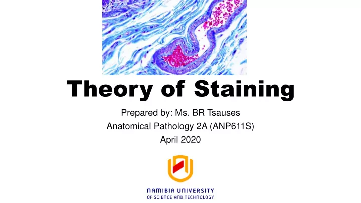

Theory of Staining Prepared by: Ms. BR Tsauses Anatomical Pathology 2A (ANP611S) April 2020
Learning objectives • Understand the aims of staining • Describe why sections need to be coloured with dyes • Describe how dye bind to tissues • Understand the different types of staining • Describe the principle of the following stains; H&E Stain and PAP stain • Describe the reasons for mounting tissue
Pre-learning Quiz • Please take the pre-learning quiz before proceeding with the presentation.
Types of staining • Non-vital stains – staining of dead tissue that has been fixed, processed and sectioned • Vital stains – the colouring of living tissue/cells either using very dilute dyes or by the phagocytic action of macrophages ingesting dye particles. • Histochemical stains - utilizes a true chemical reaction in the tissue and matches what would happen if the reaction was performed in a test tube.
Types of staining cont … • Lysochrome – the staining of neutral lipids/fats whereby elective solubility allows the dye molecules to leave the solvent and enter the lipid. • Silver Impregnation – Depositing metals onto or into tissue components. Silver is the metal most commonly employed, • Injection – the introduction of a coloured compound into the tissue to highlight various structures
Types of staining cont … • Fluorochrome – staining is affected by combining a fluorochrome with a tissue entity, which is visualized under fluorescent light. • Immunostaining – staining is based on an antibody-antigen reaction whereby a labelled antibody permits the site of the reaction to be visualised.
Why are stains taken into the tissue • Stain uptake due to dye-tissue or reagent-tissue affinities • Affinity – the tendency of a stain to transfer from solution onto section. • Affinity also describes attractive forces thought to bind dye to tissue. • The affinity’s magnitude depends on every factor favouring or hindering the movement
Staining mechanisms • Ionic bonds • Hydrogen bonds • Van der Waals forces • Covalent bonds • Hydrophobic interactions
Ionic bonding / Coulombic attractions • Ionic bonding involves electrostatic attraction between oppositely charged ions • Anionic (negatively charged) dyes will bind to cations (positively charged) in the tissue.
Ionic bonding stains • Negatively charged eosin ions will stain positively charged tissue • Acidophilic – any tissue component that stain with an acid dye • Eosinophilic – tissue components staining with eosin, e.g collagen, red blood cells and the cytoplasm of many cells. • Positively charged methylene blue ions will stain negatively charged tissue ions • Anionic dyes are also called acid dyes in histology because they are derived from coloured acids.
Ionic bonding stains cont …. • Binding of dyes depends on tissue ionization • Acid pH levels favour staining with anionic dyes • Alkaline conditions favour staining with cationic dyes • Salt concentrations affect dye staining
Hydrogen bonding • Hydrogen bonding is a dye-tissue attraction arising when a hydrogen atom lies between two electronegative atoms (e.g oxygen and nitrogen) . • Hydrogen bonding is not affected by pH or salt concentration but is affected by strong hydrogen-bonding agents e.g water • Staining of amyloid by Congo red stain uses hydrogen -bond staining
Van der Waals forces • Van der Waals forces are short-range forces and will only have an effect if the two atoms are between 0.12 and 0.2nm apart. • These are electrostatic attractions that always exist between the electrons of one atom and the nucleus of another. • Intermolecular attractions such as dipole-dipole, dipole-induced dipole and dispersion forces
Van der Waals forces cont …. • A dipole is a molecule in which the electrons are unsymmetrically distributed, so that one end carries a fractional electrical charge relative to the other end, e.g water is dipole. • Van der Waals forces occur when the surface shape of the tissue protein and the shape of the dye match, then bonds are formed.
Van der Waals forces cont … • The adhesion of the section to the slide involves van der Waals interactions between the section and the glass. • Van der Waals bonds are unaffected by pH, ions and hydrogen - boding agents.
Covalent bonds • Covalent bonding between tissue and stain involves polar covalent bonds between metal ions and mordant dyes.
Hydrophobic interaction •Hydrophobic bonds hold dyes in tissues by the exclusion of water from the regions of hydrophobic groups. • This is the tendency of hydrophobic groups to come together, even though initially dispersed in an aqueous environment. •Hydrophobic interactions are unaffected by hydrogen - bonding agents or salts •Hydrophobic interactions play a major role in the staining of lipids.
General structure of dye molecules • A dye molecule have two parts; chromophore and auxochrome • Chromophores – (Colour bearer) any compound that makes an organic compound coloured e.g Quininoid, azo and nitro groups • Auxochromes - (Ionizing group) an ionizing group that permits a dye to bind to tissue.
General structure of dye molecules cont … • The common auxochromes used in histology are hydroxyl (OH), Methly (CH3) and amino (NH2) groups. Auxochromes also intensify the colour • Fluorochrome – absorbs ultraviolet. Violet, blue or green light and emits light of longer wave length. Fluorochrome compounds are used in fluorescence microscopy.
The coloured index (C.I) • The standard list of all dyes, containing their synonyms and their structures • Each dye is given an individual number and listed along with its names and properties. • E.g Eosin Y (C.I. 45380) • CI numbers are arranged according to their structures; Nitro dyes, Azo dyes, Triarly methane dyes, Anthraquinone, Xanthene and Thiazine
Factors affecting Dye binding • pH • temperature • concentration • salt content • fixative (formalin react with the NH2 group, because this is the primary group for binding eosin, tissue fixed in formalin will bind less eosin than when fixed in some of the other solutions.
Histology classification of dyes • Histology classification is based on the dyes action on the tissue • Basic dyes – these are cationic dyes and will stain anionic or acidic materials (e.g the phosphates in nucleic acid) in the the tissue. • Nuclear stains contain basic dyes • Acidic substances that stain with basic dyes are termed basophilic
Histology classification of dyes cont … • Acidic dyes - these are anionic dyes and will stain cationic or basic groups in tissue such as amino groups. • Acidic dyes stain proteins in the cytoplasm and connective tissue. • Substances that stain with acid dyes are called acidophilic
Histology classification of dyes cont …. • Neutral Dyes- simply compounds of basic and acidic dyes. Such dye complexes will stain both nucleus and cytoplasm, e.g Romanowsky stains • Amphoteric dyes - have both anionic and cationic groups, but on the same ion. Such dyes stain either the nucleus or the cytoplasm if conditions are appropriate. • Natural dyes – dye substances extracted from natural sources.
Basophilic & Eosinophilic Staining • Acid components of a cell (like nuclear chromatin or chromosomes) stain with basic dyes (e.g., hematoxylin) and these components are referred to as basophilic or hematoxylinophilic (blue). • Basic components (various kinds of cytoplasm and intercellular substance) take acid dyes (e.g., eosin), and are called acidophilic or eosinophilic (pink).
Basophilic & Eosinophilic Staining (example)
Non-dye constituents of staining solutions • Mordants – non-dying compound to improve the binding of the dye • Trapping agents – always applied after the dye to prevent the escape of dye that has entered the tissue entity, e.g the Gram’s stain uses iodine as a trapping agent to form large aggregates with crystal violet.
Non-dye constituents of staining solutions •Accentuators and accelerators – these are materials added to staining solutions to improve the staining reaction. • Accentuators control pH and accelerators their mechanisms of enhancement is not known.
Metachromatic dyes • Metachromasia – when a dye stains a tissue component a different colour to the dye solution. • Toluidine blue is a strong basic blue dye that stain nuclei a deep blue colour and mast cell granules a pink colour.
Important dyes in histology Nuclear stains • Haematoxylin • Carmine and Carminic acid • Methylene blue • Neutral Red and Safranine O • Methlyl green • Toluidine blue
Cytoplasmic stains • Eosin • Methyl blue and aniline blue • Fast green FCF and Light green SF • Orange G, metanil yellow and martius yellow
Nuclear stains • The nucleus of a eukaryotic cell contains the two nucleic acids. • The dye used as nuclear stains impart colour to the chromatin by dinding to the nucleic acid, the nucleoprotein or both substances • Cationic dyes are used to stain the nucleus • The aqueous solution of the dye is usually acidified by addition of acetic acid or suitable buffer.
Recommend
More recommend