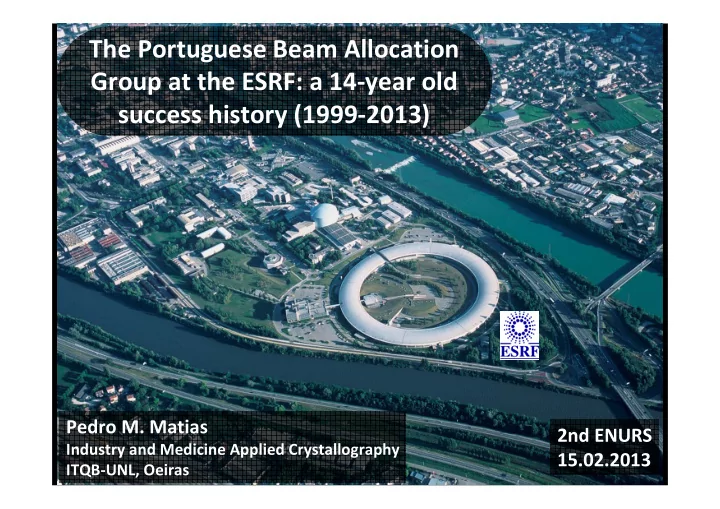

The Portuguese Beam Allocation Group at the ESRF: a 14 ‐ year old success history (1999 ‐ 2013) Pedro M. Matias 2nd ENURS Industry and Medicine Applied Crystallography 15.02.2013 ITQB ‐ UNL, Oeiras
The ESRF at a glance Founded in 1998 Began operation in 1994 Annual budget: ca. 80 M€ Mem bers (minimum 4% shares, full voting rights) : France, Germany, Italy, United Kingdom, Spain, Switzerland, Benesync (Belgium & The Netherlands), Nordsync (Denmark, Finland, Norway, Sweden) Scientific Associates (< 4% shares, limited voting rights) : Austria, Portugal , Israel, Poland, Centralsync (Czech Republic, Hungary, Slovakia).
Portugal, MX and the ESRF The first Portuguese MX users of the ESRF were given access through a collaboration with the EMBL Grenoble Outstation in 1995-6 Portugal joined the ESRF as a scientific associate in 1997 (1% ). Novel statute created to accommodate the Portuguese membership, later allowed other small European countries to join the ESRF. In 1999 the MX BAG scheme was created to promote a more efficient and productive use of the beamlines: 1 shift = 8 hours = MANY experim ents In recognition of their excellent work, the Portuguese MX groups were invited to form one of the first BAGs Today, BAG use of the ESRF MX beamlines accounts for more than 90% of the available beamtime
The Portuguese MX BAG in 1999 I TQB – Universidade Nova de Lisboa Maria Arménia Carrondo REQUI MTE/ FCT – Universidade Nova de Lisboa Maria João Romão I BMC – Universidade do Porto Ana Margarida Damas
The Portuguese MX BAG in 2013 I TQB – Universidade Nova de Lisboa Margarida Archer Maria Arménia Carrondo Carlos Frazão Pedro Matias I GC – Fundação Calouste Gulbenkian Alekos Athanasiadis REQUI MTE/ FCT – Universidade Nova de Lisboa Maria João Romão I BMC/ I MEB - Porto Luís Gales Pinto Sandra Macedo-Ribeiro João Morais-Cabral Pedro Pereira
The BAG scheme in Practice Beam allocation every 6 m onths Yearly Report : alternating Progress Report and Full Report Report evaluation by Review Panel determines maintenance of BAG status and overall beam allocation Our scores have been “good”; To improve them to “excellent” in order to increase beam allocation, we need: - Work in more challenging projects (e.g., membrane proteins) - More publications in high IF journals (e.g., Science, Nature, etc.)
An Overview of the ESRF Source: http://www.esrf.fr/AboutUs/GuidedTour/Animation
Source: http://www.esrf.fr/AboutUs/GuidedTour/Animation
The MX beamlines at ESRF Storage Ring Optics cabin Sample to study Experimental cabin Control cabin Source: http://www.esrf.fr/AboutUs/GuidedTour/Animation
The MX beamlines at the ESRF
Source: http://www.embl.fr/services/synchrotron_access/id14-4/
100 μ m
BAG statistics – shifts used 100 87 85 90 80 66 70 Shifts used 60 57 60 49 50 40 30 20 10 0 2001 ‐ 2003 1 2003 ‐ 2005 2 2005 ‐ 2007 3 2007 ‐ 2009 4 2009 ‐ 2011 5 2011 ‐ 2013 6
BAG statistics – PDB depositions 40 35 35 28 28 30 PDB entries 25 20 15 13 13 10 5 0 1 2 3 4 5 2001 ‐ 2003 2007 ‐ 2009 2009 ‐ 2011 2003 ‐ 2005 2005 ‐ 2007
BAG statistics - Publications 20 15 15 13 13 13 Publications 12 11 11 10 10 10 10 9 8 6 6 5 1 0 2001 ‐ 2003 2007 ‐ 2009 2009 ‐ 2011 2003 ‐ 2005 2005 ‐ 2007 1 2 3 4 5 Crystallization Reports Journals with IF > 4 Journals with IF < 4
Publications with IF>4: 2001-2011 10 15 0 5 N a t u r e 1 1 S c i e n c e 2 1 N a t u r e S B 3 2 A c t a D 4 9 J A C S 5 3 P N A S 6 4 S t r u c t u r e 7 2 J B C 8 13 P L o S O N E 9 4 J M B 10 7
Novel anticoagulant mechanism SerRS, the main player in C. albicans in the malaria mosquito genetic code alteration - Anophelin binds to thrombin in the - Crystal structures of the two natural reverse direction of bona fide isoforms of Candida albicans seryl- substrates disrupting the catalytic tRNA synthetase (SerRS-Leu / SerRS- triad Ser) - Its compact size and resistance to - Ambiguous codon localization proteolysis might the design of novel tailored to minimize protein antithrombotics misfolding events Figueiredo et al . (2012) Proc. Natl. Acad. Sci. USA 109 , E3649-58 Rocha et al . (2011) Proc. Natl. Acad. Sci. USA 108 , 14091-6
Main Research lines & Highlights 1 ‐ Molybdopterin Enzymes • Aldehyde Oxidases: The first mammalian aldehyde oxidase structures (mouse and human) • Detailed mechanistic studies and novel mechanisms based on atomic ‐ resolution structures (MOP, NAP) 2 ‐ Drug Design • CO Releasing Molecules (CORM) binding mechanism to plasma proteins • Adducts of Human Transferrin and Vanadium as anti ‐ diabetic agents • Design of potent and specific inhibitors of proteases based on Trypsin studies 3 ‐ Cellulosome: a megaDalton complex for cellulose degradation: • Carbohydrate binding Modules • Glycoside hydrolases • Cohesin ‐ dockerin complexes
Molybdopterin Enzymes • Detailed mechanistic studies • Ligand identification and novel mechanisms Ethylene ‐ glycol & Glycerol inhibited AOR Oxidized & Reduced Nitrate Reductase @1.5 Å @1.7 Å O? S Reduced RS SR RS SR RS SR Mo VI Mo V Mo IV • First structural evidence for a Mo ‐ C bond in a Mo and ligand ‐ based redox chemistry biological system • Possible to distinguish Mo ‐ O and Mo ‐ C bonds • New mechanism for nitrate reduction: • Substrates may also bind to the protein by a η 2 Ligand ‐ based redox chemistry coordination • Unanticipated sulfur ligand • Partial disulfide bond Romão, Dalton Trans, 2009 Santos ‐ Silva et al, JACS , 2009 Coelho et al , J Mol Biol, 2011 Correia et al, submitted POSTER & Oral communication
Molybdopterin Enzymes • First Crystal Structures of mammalian Aldehyde Oxidases • Identification of new metallic clusters E. coli Periplasmic Aldehyde Oxidase 1.8 Å Mouse Aldehyde Oxidase 3.0 Å @ SLS @ ID14 ‐ 1 & ID23 ‐ 1 Extremely poor crystals and weak diffraction! Identified a new (P1 (4 mols/au; 1300 aa)) [4Fe ‐ 4S] cluster! • Insights into substrate specificity • Important in drug metabolism & increasing importance in recent drug design programs (Pfizer) • The first monomeric XO ‐ related enzyme Coelho et al, Drug Metab Dispos. 2011 • Unique member of the XO family POSTER Coelho et al, J Biol. Chem. 2012
Drug Design • CO Releasing Molecules (CORM) binding mechanism to plasma proteins • Vanadium – Transferrin adducts • Trypsin – Inhibitors binding Trypsin and inhibitors of Urokinase 1.5 Å CORM – Lysozyme adducts 1.7 Å @ ID29 @ ID14 ‐ 1 & ID23 ‐ 1 • Identification of inhibitors binding site • Covalent bond B ‐ Ser195 Spencer et al, to be submitted Vanadium ‐ Human Transferrin adducts • Identification of the binding sites of V compounds • Structural characterization of protein – CORM interactions • Conformational changes upon binding (SAXS) • Insights into CO release and polyoxometallate formation • Urea Gel Electrophorese to control protein modifications upon binding Santos ‐ Silva et al, JACS, 2011; Curr Med Chem, 2011 Santos et al , JIB, 2012 POSTER Mehtab et al, J Inorg Biochem , in press Seixas et al, Dalton T, 2012
Cellulosome : a megaDalton complex for cellulose degradation Type II Cohesin ‐ Dockerin complex Crystal structures of the type II interaction in Clostridium Family 124 Glycoside thermocellum Hydrolase Carvalho et al, JMB , 2005 First structure of a Alves et al, in prep representative member POSTER & Oral communication Brás et al, PNAS , 2011 Type I Cohesin ‐ Dockerin complex First crystal structure of a type I interaction Carvalho et al, PNAS , 2003 Carbohydrate Binding Modules Carvalho et al, PNAS , 2007 types B and C Brás et al, JBC , 2012 Carvalho et al, JBC , 2004 POSTER Najmudin et al, JBC, 2006 Viegas et al, FEBS J , 2008 Santos ‐ Silva et al, BBA , 2010
Cyprinid Herpes Virus 3 Orf122 Crystal Structure ‐ A poxvirus ‐ like Zalpha domain ID23 ‐ 1 Unlike other Zalpha domains, ORF112 forms a dimer through a unique domain ‐ swapping mechanism. Tomé AR, Ku ś K., Correia S., Paulo L., Zacarias S., de Rosa M., Figueiredo D., Parkhouse RME. and Athanasiadis A. (2013) J. Virology in print
Structure Characterization of a multicopper oxidase from Campylobacter jejuni CGUG11284 Campylobacter jejuni is a Pathogenic, Gram ‐ negative bacterium, that is the most common cause of human gastroenteritis and bacterial food poisoning. McoC – is a periplasmic multicopper oxidase thought to be involved in copper homeostasis. The McoC structure displays a characteristic laccase ‐ like fold, with three cupredoxin domains and two types of copper centres: a T1 copper centre in domain III and a tri ‐ nuclear center, with two T3 and one T2 copper atoms, localised Resolution 1.95 Å between domain I and III. C. Silva et.al. Metallomics , 2012;4(1):37 ‐ 47.
Recommend
More recommend