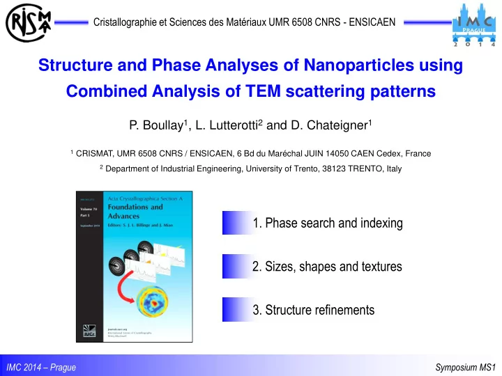

Cristallographie et Sciences des Matériaux UMR 6508 CNRS - ENSICAEN Structure and Phase Analyses of Nanoparticles using Combined Analysis of TEM scattering patterns P. Boullay 1 , L. Lutterotti 2 and D. Chateigner 1 1 CRISMAT, UMR 6508 CNRS / ENSICAEN, 6 Bd du Maréchal JUIN 14050 CAEN Cedex, France 2 Department of Industrial Engineering, University of Trento, 38123 TRENTO, Italy 1. Phase search and indexing 2. Sizes, shapes and textures 3. Structure refinements IMC 2014 – Prague Symposium MS1
EPD Quantitative Analysis of Electron Powder Diffraction powder single-crystal ◄ tens of micrometer ► XRD cell, symmetry and structure Mo K =0,7107 Å phase S/M, structure and microstructure (size, shape, texture) PEDT ◄ tens of nanometer ► Nanoparticles (NP) 200kV =0,0251 Å Precession Electron Diffraction Tomography Electron Powder Diffraction (EDP) patterns IMC 2014 – Prague Symposium MS1
EPD Quantitative Analysis of Electron Powder Diffraction Quantitative and statistically representative analysis of crystallite sizes and shapes, structure and crystallographic texture of nanoparticles in the form of powders and thin films? Extraction of intensities from electron diffraction “ring patterns” for quantitative or semi- quantitative analysis … Vainshtein (1964), … PCED 2.0 : X.Z. Li, Ultramicroscopy 110 (2010) 297-304 ProcessDiffraction : J.L. Labar, Microsc. Microanal. 15 (2009) 20-29 TextPat : P. Oleynikov, S. Hovmoller and X.D. Zou in Electron Crystallography L. Lutterotti Nuclear Inst. and Methods in Physics Res. B268 (2010) 334-340. The MAUD program : IMC 2014 – Prague Symposium MS1
MAUD Materials Analysis Using Diffraction http://www.ing.unitn.it/~maud/ MAUD Rietveld pattern fitting Delft size-strain (PV) Popa anisotropic Evolutionary Size/Strain distributions Simulated Annealing Planar faulting (Warren) Marquardt (Least squares) Turbostratic (Ufer) Metadynamics optimization Size-Strain Indexing Simplex (Nelder-Mead) Genetic (COD phase search procedure) March-Dollase Peak location X-ray Harmonic Texture Neutron (E)WIMV Standard Functions Electron Peak fitting Residual stresses Geometric Structure refinement Voigt, Reuss, Hill Triaxial Stress IMC 2014 – Prague Symposium MS1
MAUD EPD Intensity extraction Intensity extraction along the rings by segments using an ImageJ plugin pixel size ► pixels to mm 120 patterns 2D plot estimation of the center position using a reference circle 3° on the screen CAKING chi=phi=0° / omega=90° / eta: 0° to 360° calibrate the distance specimen/detector ► mm to 2 q IMC 2014 – Prague Symposium MS1
EPD Line Broadening in Powder Diffraction 1D XRPD-like pattern (360° summed intensity) peak location and intensities peak broadening vs. d hkl b(x): background => pic at 0° + polynomial function 2 q (°) => Q (Å -1 ) measured profile h(x) = f(x) g(x) + b(x) IMC 2014 – Prague Symposium MS1
EPD Line Broadening in Powder Diffraction h(x) = f(x) g(x) + b(x) Line broadening causes sample contribution instrumental broadening • instrumental broadening • finite size of the crystals (acts like a Fourier truncation: size broadening) • imperfection of the periodicity (due to d h variations inside crystals: microstrain effect) • generally: 0D, 1D, 2D, 3D defects All quantities are average values over the probed volume ► electrons, x-rays, neutrons: complementary ► distributions: mean values depend on distributions’ shapes Extraction of f(x) can be obtained by a whole-pattern (Rietveld) analysis Need to know g(x) the instrumental broadening ! L. Lutterotti and P. Scardi, J. of Appl. Crystallogr. 23, 246-252 (1990) The instrumental Peak Shape Function is obtained by analysing nanoparticules of known sizes and shapes as obtained from X-ray analyses IMC 2014 – Prague Symposium MS1
EPD EPD vs XRPD Mn 3 O 4 hausmannite ( L. Sicard et al, J. Magn. Magn. Mater. 322 (2010) 2634-2640 ) Bruker D8 / Lynx Eye 1D g(x) ► f(x) structure =1.54056 Å (Cu K 1 ) SG: I 4 1 /a m d a=5.764(2)Å and c=9.448(4)Å 64 Å POPA 53 Å anisotropic shape TOPCON 2B / CCD ORIUS pattern matching f(x) ► g(x) =0.0251Å background substracted a=5.7757(2)Å and c=9.4425(4)Å IMC 2014 – Prague Symposium MS1
EPD Sizes, shapes and textures Microstructure of nanocrystalline materials: TiO 2 rutile (1) from phase search: TiO2 rutile P4 2 /mnm a= 4.592Å a=2.957Å (COD database ID n°9001681) 72 patterns 5° 20nm FEI Tecnai / CCD USC1000 / =0.0197Å (1) M. Reddy et al., ElectroChem. Com. 8 (2006) 1299-1303 IMC 2014 – Prague Symposium MS1
EPD Sizes, shapes and textures 4-circles diffract. / INEL CPS structure RX = CuK average anisotropic crystallite size pattern matching EPD IMC 2014 – Prague Symposium MS1
EPD Sizes, shapes and textures 6 μ m 0.5 μ m decreasing the selected area Texture :: intensity variation along the rings fit fit no texture E-WIMV data data Q (Å -1 ) Q (Å -1 ) IMC 2014 – Prague Symposium MS1
EPD Sizes, shapes and textures The features available in MAUD allow a full quantitative texture analysis for general cases (not only fiber textures) from EPD patterns with the obtention of accurate pole figures QTA analysis of Pt thin film deposited on Si {111} pole figure from ODF refinement +25° to -25° step 5° For application on textured thin film see also M. Gemmi et al., J. Appl. Cryst. 44 (2011) IMC 2014 – Prague Symposium MS1
EPD Structure refinement ► microstructural features can be obtained in the pattern-matching mode ► not convincing using structure factors from kinematical approximation … … much better when using the 2 -beam or Blackman correction TEM =0.0251 Å CuK (150 o 2 q ) √I*Q=f(Q) I=f(Q) NP TiO 2 rutile synchrotron =0.486 Å Pair Distribution Function analyses on EPD ▼ A.M.M. Abeykoon, C.D. Malliakas, P. Juhás, E.S. Božin , M.G. Kanatzidis, S.J.L. Billinge, Z. Kristallogr. 227 (2012) 248 IMC 2014 – Prague Symposium MS1
EPD Structure refinement ► microstructural features can be obtained in the pattern-matching mode ► not convincing using structure factors from kinematical approximation … … much better when using the 2 -beam or Blackman correction Rietveld refinement :: combined analysis √I*Q=f(Q) NP TiO 2 rutile 5.0 10.0 IMC 2014 – Prague Symposium MS1
EPD Quantitative Analysis of Electron Powder Diffraction Structure and Phase Analyses of Nanoparticles using Combined Analysis of TEM scattering patterns automatic phase search procedure ( COD database, multi-phases ) average lattice cell parameters and crystallite size ( anisotropic shapes ) accurate texture analysis ( general cases, ODF, … ) whole-pattern S/M procedure … can be obtained in the Pattern matching mode ( kinematical approximation ) structure determination and refinement are possible within MAUD … implementation of PDF approach soon http://nanoair.dii.unitn.it:8080/sfpm and http://cod.iutcaen.unicaen.fr IMC 2014 – Prague Symposium MS1
EPD Quantitative Analysis of Electron Powder Diffraction Structure and Phase Analyses of Nanoparticles using Combined Analysis of TEM scattering patterns automatic phase search procedure ( COD database, multi-phases ) average lattice cell parameters and crystallite size ( anisotropic shapes ) accurate texture analysis ( general cases, ODF, … ) … can be obtained in the Pattern matching mode structure refinements are possible within MAUD ( kinematic or Blackman ) … implementation of PDF approach soon Thank you for your attention V. Pralong and V. Caignaert (TiO 2 nanoparticules) @ CRISMAT – Caen L. Sicard and S. Ammar (Mn 3 O 4 nanoparticules) @ ITODYS – Paris 7 S. Gascoin (XRD measurements) @ CRISMAT – Caen ANR FURNACE, BAMBI IMC 2014 – Prague Symposium MS1
Recommend
More recommend